


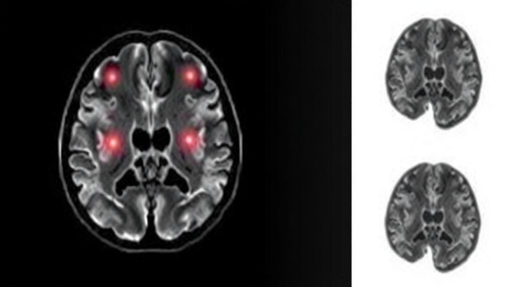
Every surgeon who operates on the pituitary gland faces the same nerve-racking question moments before the first instrument touches the tumor: will it come out like soft putty, or will it resist like rubber? The answer shapes everything, from the surgical approach to the likelihood of a complete removal. Now a team of researchers in China has shown that a mathematical model borrowed from the physics of random movement can answer that question in advance, using nothing more than a specially tuned magnetic resonance imaging scan. The study, published in BMC Medical Imaging, suggests that the way water molecules wander through tumor tissue encodes a hidden signature of its texture and of how much of it a surgeon can realistically expect to remove.
The research, led by Chun-Qiu Su, Hong-Li Bian and Shan-Shan Lu of the First Affiliated Hospital with Nanjing Medical University, together with colleagues in neurosurgery, pathology and industry, focused on pituitary adenomas, benign tumors arising in the pea-sized gland at the base of the brain. Although these growths are not cancerous, their location makes them treacherous. They press on the optic nerves, disrupt hormone production and often extend sideways into the cavernous sinuses, blood-filled channels that carry major arteries and nerves. The standard treatment is endoscopic transsphenoidal surgery, in which instruments are threaded through the nose and sphenoid sinus to reach the tumor from below. How well that operation goes depends heavily on one physical property: consistency.
Soft tumors can be suctioned, curetted and peeled away from surrounding structures with relative ease, allowing surgeons to achieve what is called a high extent of resection, or EOR. Hard tumors, by contrast, are fibrous and stubborn. They cling to the optic apparatus and the cavernous sinus, and surgeons must balance the goal of complete removal against the risk of devastating complications such as permanent vision loss or arterial injury. Preoperative imaging has long been asked to predict consistency, but conventional techniques have struggled. The workhorse measurement, the apparent diffusion coefficient or ADC derived from diffusion-weighted MRI, offers only a single averaged number that blurs together many different microscopic environments inside a tumor.
The Nanjing team turned instead to a richer mathematical framework known as the continuous-time random-walk, or CTRW, model. Classical diffusion theory, rooted in Einstein’s description of Brownian motion, assumes that particles take small, evenly timed steps in a random walk, producing the familiar Gaussian spreading of a drop of ink in still water. Biological tissue rarely behaves so politely. Water molecules in a tumor encounter cell membranes, collagen fibers, pockets of extracellular matrix and regions of differing cell density, so their movement becomes anomalous: steps are not uniform in length or in time. The CTRW model embraces this messiness by introducing parameters that quantify exactly how the walk departs from idealized Brownian motion.
Three parameters carry the diagnostic weight. The anomalous diffusion coefficient, written D(m), reflects the overall mobility of water in the tissue. The temporal heterogeneity parameter, alpha, describes how waiting times between molecular jumps are distributed; values below one indicate that motion is slower and more trapped than normal diffusion would allow. The spatial heterogeneity parameter, beta, characterizes how step lengths deviate from the Gaussian ideal, with lower values signaling heavier-tailed distributions and more obstructed pathways. By acquiring diffusion MRI at multiple so-called b-values, which progressively sensitize the scan to molecular displacement, the researchers could fit these parameters to the signal decay measured across each whole tumor.
To test whether these numbers mean anything clinically, the team enrolled 53 patients with pituitary adenomas in a prospective study. Each patient underwent multi-b-value diffusion MRI before surgery, and the CTRW parameters were calculated from regions of interest covering the entire tumor. Then came the ground truth: during the operation, the neurosurgeons classified each tumor as soft or hard based on what their instruments actually encountered. The results were striking. Hard tumors showed significantly lower alpha values than soft ones, 0.472 versus 0.649 on average, and significantly lower beta values as well, 0.752 versus 0.806. The anomalous diffusion coefficient and the conventional ADC were also reduced in hard tumors, though with weaker statistical support.
The pathology added a satisfying mechanistic layer. Tumor hardness in pituitary adenomas is largely a story of collagen, the fibrous structural protein that stiffens tissue the way it stiffens skin or tendon. When the researchers correlated collagen content measured in surgical specimens with the imaging parameters, they found a significant negative relationship with every diffusion measure, and the temporal heterogeneity parameter alpha showed the strongest association of all, with a correlation coefficient of minus 0.492. In other words, the more collagen packed the tumor, the more constrained and anomalous the molecular walk became, and the lower alpha fell. The physics was reading the biology directly.
Consistency, however, is only half the story; what patients and surgeons ultimately care about is how much tumor can be safely removed. When the team analyzed which factors predicted an extent of resection greater than 95 percent, patients who achieved that benchmark had significantly higher alpha and beta values and lower Knosp grades, the standard radiological scale for how far a tumor invades the cavernous sinus. In multivariable logistic regression, alpha remained an independent predictor, with each 0.1 increase raising the odds of extensive removal by 10 percent, while beta carried an even stronger effect, with each 0.1 increase raising the odds by 20 percent. Knosp grade also mattered independently, with each higher grade cutting the odds by roughly 71 percent, a reminder that anatomical invasion still governs surgical feasibility.
The most compelling result emerged when the pieces were combined. A model incorporating Knosp grade together with alpha and beta achieved an area under the receiver operating characteristic curve of 0.941, a level of discrimination that approaches clinical usefulness, and it significantly outperformed Knosp grade alone. This matters because the Knosp grade, however valuable, says nothing about tissue texture; a tumor that spares the cavernous sinus can still be fibrous and difficult, and one that invades it can still be soft and yielding. Adding the CTRW parameters gives the surgical team a two-dimensional picture: where the tumor has gone and what it will feel like when they get there.
The implications extend beyond the operating theater. Preoperative knowledge of tumor consistency could influence surgical planning, patient counseling and even the choice between surgical strategies, while the CTRW framework itself, already being explored in other tumor types, offers a general-purpose lens on tissue microstructure that goes far beyond what a single ADC value can deliver. The study has limitations worth noting: 53 patients is a modest sample, the soft-versus-hard classification rests on intraoperative judgment, and the findings await external validation in independent cohorts. But the core message is hard to dismiss. Water molecules jostling through a pituitary tumor carry information about collagen, texture and resectability, and with the right physics, an ordinary MRI scanner can be taught to listen. For patients facing surgery at the crossroads of the skull base, that listening could soon translate into better-informed decisions and safer operations.
Subject of Research: Continuous-time random-walk diffusion MRI for predicting tumor consistency and extent of resection in pituitary adenomas
Article Title: Continuous-time random-walk diffusion model for predicting tumor consistency and extent of resection in patients with pituitary adenomas
Article References: Su, C.-Q., Bian, H.-L., Wang, B.-B., Tao, C., Pan, M.-H., Tang, R., He, Y.-X., Liu, D., & Lu, S.-S. (2026). Continuous-time random-walk diffusion model for predicting tumor consistency and extent of resection in patients with pituitary adenomas. BMC Medical Imaging. https://doi.org/10.1186/s12880-026-02820-3
Image Credits: AI Generated
DOI: 10.1186/s12880-026-02820-3
Keywords: pituitary adenoma, continuous-time random-walk, diffusion MRI, tumor consistency, extent of resection, apparent diffusion coefficient, collagen, Knosp grade, transsphenoidal surgery, anomalous diffusion, BMC Medical Imaging, preoperative imaging
Cite Scienmag News
Ophelia Keating. (October 8, 2026). Random-Walk MRI Physics Predicts How Hard a Brain Tumor Is Before Surgery. Scienmag. https://scienmag.com/random-walk-mri-physics-predicts-how-hard-a-brain-tumor-is-before-surgery/
Ophelia Keating. "Random-Walk MRI Physics Predicts How Hard a Brain Tumor Is Before Surgery." Scienmag, 8 October 2026, https://scienmag.com/random-walk-mri-physics-predicts-how-hard-a-brain-tumor-is-before-surgery/. Accessed 8 October 2026.
Ophelia Keating. "Random-Walk MRI Physics Predicts How Hard a Brain Tumor Is Before Surgery." Scienmag. October 8, 2026. https://scienmag.com/random-walk-mri-physics-predicts-how-hard-a-brain-tumor-is-before-surgery/


