


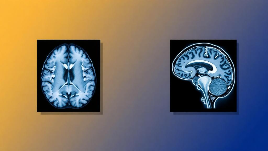
Positron emission tomography is one of the most powerful tools in modern medicine, letting physicians watch metabolism unfold inside the living brain. But the photons that PET detectors count do not travel unimpeded through the head. Bone, soft tissue, and even air absorb and scatter them, and unless that attenuation is corrected, the resulting images misrepresent how much radiotracer has actually accumulated in each region. On combined PET/CT scanners this correction is straightforward, because the computed tomography component directly measures tissue density. On integrated PET/MRI scanners, however, the magnetic resonance images that come bundled with the PET data contain no information about X-ray attenuation at all. Magnetic resonance signal intensity reflects proton relaxation behavior, not electron density, so the very scanner designed to eliminate radiation exposure from CT leaves clinicians without an obvious way to correct one of PET’s most important physical distortions.
This long-standing gap is the target of a new study published in Medical & Biological Engineering & Computing by Seyede Sajede Mousavi Eshkiki and Alireza Sadremomtaz of the University of Guilan in Iran. Rather than proposing yet another neural-network architecture, the pair set out to build and rigorously evaluate a complete, two-stage workflow that takes ordinary T1-weighted MRI of the brain and produces both a CT-like image and a fully attenuation-corrected PET scan. Crucially, they wrapped the entire pipeline in a graphical user interface, so that the deep learning machinery can be operated through inference without requiring users to write code. The work is an exercise in systems engineering as much as machine learning: the question was not whether a clever model could be designed, but whether a sequence of established components could be assembled into a workflow that behaves reliably enough to matter clinically.
The researchers drew their data from the CERMEP-IDB-MRXFDG cohort, a publicly available database of thirty-seven healthy adults who underwent fluorodeoxyglucose PET, T1-weighted and FLAIR MRI, and CT imaging of the brain. Because every subject had a real CT scan, the team could train their networks against genuine ground truth and then measure how closely the synthetic versions matched reality. They used subject-level five-fold cross-validation, meaning that in each round of testing the network had never seen the individual whose images it was correcting. This patient-level separation matters, because it prevents the model from effectively memorizing a person’s anatomy and inflating its apparent accuracy.
The first stage of the workflow is a two-dimensional U-Net, the workhorse convolutional network of biomedical image analysis, first introduced in 2015 and still favored for its efficiency and strong performance on relatively small datasets. Fed only a T1-weighted MRI volume, the network predicts a pseudo-CT: a synthetic image in Hounsfield units that approximates what a CT scanner would have measured for the same head. Once generated, this pseudo-CT is converted into an approximate linear attenuation coefficient map, the so-called mu-map that describes how strongly tissue attenuates the 511-keV photons emitted during positron annihilation. That mu-map, paired with the non-attenuation-corrected PET image, then serves as the input to a second U-Net, which synthesizes a predicted attenuation-corrected PET scan directly.
The logic of this second stage is subtle and worth unpacking. In principle, one could simply apply the mu-map mathematically to correct the PET data, as conventional reconstruction does. But the second network learns the mapping from uncorrected PET plus attenuation information to corrected PET, allowing it to absorb residual imperfections in the pseudo-CT. If the synthetic attenuation map slightly misestimates bone density, for example, the second network can still learn to produce an output consistent with properly corrected images, because it has seen many paired examples of what uncorrected and corrected PET look like for the same brain. The two networks therefore act as a chain, with the first converting MRI into attenuation-relevant anatomy and the second converting that anatomy plus raw PET into a quantitatively usable scan.
The performance figures tell a encouraging story with a familiar caveat. The pseudo-CT model achieved a whole-image mean absolute error of 72.26 plus or minus 20.11 Hounsfield units and a structural similarity index of 0.881 plus or minus 0.024 against the real CT scans. The authors note that the residual error was concentrated in bone-dominated regions, which is exactly where MRI-to-CT synthesis has always struggled. Bone produces almost no signal on conventional MRI, so the network must essentially infer cortical skeleton from the shape of the brain and surrounding tissues. Decades of attenuation-correction research, from segmentation-based methods to atlas-based approaches and zero-echo-time MRI sequences, have wrestled with the same limitation, and deep learning approaches inherit it to varying degrees.
Where the workflow truly shines is in the PET correction itself. Compared with the raw, non-attenuation-corrected PET images, the predicted attenuation-corrected PET achieved a 76.02 percent lower normalized mean absolute error and a 73.33 percent lower normalized root mean square error relative to the reference attenuation-corrected PET. In other words, the two-stage pipeline recovered roughly three quarters of the quantitative error introduced by ignoring attenuation. For brain imaging, where attenuation is less severe than in the thorax or abdomen but still clinically significant, that level of recovery brings synthetic correction into the same conversation as established vendor solutions.
The exploratory standardized uptake value analysis adds a dose of realism. Standardized uptake values are the currency of quantitative PET, and the workflow delivered a mean percentage error of 8.66 percent for SUVmean but 22.08 percent for SUVmax, with greater variability across subjects in the maximum metric. This pattern is well understood in the field: SUVmean averages over a region and tends to forgive localized errors, while SUVmax hinges on a single hottest voxel and is exquisitely sensitive to small misestimates of attenuation, particularly near bone. The authors are appropriately measured, framing these results as supporting internal feasibility on the CERMEP dataset while emphasizing that nested cross-validation and external validation on independent cohorts remain necessary before any clinical claim can be made.
What makes the study notable beyond its numbers is its orientation toward usability and transparency. The graphical user interface lowers the barrier for imaging scientists and clinicians who lack programming expertise, echoing earlier efforts such as the Deep Learning Application Engine to bring neural networks out of notebooks and into the imaging suite. Just as importantly, the team has released the source code, trained models, cross-validation fold assignments, configuration files, and evaluation outputs through the Zenodo repository, allowing other groups to reproduce the results, stress-test the workflow on their own data, and extend it. In a subfield where reproducibility has often lagged behind headline accuracy figures, that openness is a meaningful contribution in its own right.
The broader significance lies in what accessible attenuation correction could unlock. PET/MRI offers simultaneous acquisition of molecular and anatomical information with a fraction of the radiation dose of PET/CT, making it attractive for pediatric imaging, longitudinal neuroscience studies, and repeated oncological follow-up. If a workflow like this one, running on nothing more exotic than a standard T1-weighted scan, can deliver dependable attenuation correction without dedicated sequences or vendor-proprietary tools, it could lower the cost and complexity of quantitative PET/MRI for research centers worldwide. The caveats stand: thirty-seven healthy subjects, a single tracer, and no external cohort yet. But as a carefully documented proof of feasibility, the study charts a practical path from the physics problem that has shadowed PET/MRI since its inception toward a solution that any lab with a GPU and a GUI could pick up and run.
Subject of Research: Deep learning-based attenuation correction for brain PET/MRI using pseudo-CT synthesis
Article Title: A two-stage PET/MRI attenuation-correction workflow for pseudo-CT and AC PET synthesis with GUI-based inference
Article References: Eshkiki, S. S. M., & Sadremomtaz, A. (2026). A two-stage PET/MRI attenuation-correction workflow for pseudo-CT and AC PET synthesis with GUI-based inference. Medical & Biological Engineering & Computing. https://doi.org/10.1007/s11517-026-03685-y
Image Credits: AI Generated
DOI: 10.1007/s11517-026-03685-y
Keywords: PET/MRI, attenuation correction, pseudo-CT, U-Net, deep learning, mu-map, NAC PET, SUV, CERMEP-IDB-MRXFDG, medical imaging, radiotracer imaging, quantitative PET
Cite Scienmag News
Cassandra Pierce. (October 5, 2026). AI Workflow Turns Brain MRI Into CT-Like Images to Sharpen PET Scans. Scienmag. https://scienmag.com/ai-workflow-turns-brain-mri-into-ct-like-images-to-sharpen-pet-scans/
Cassandra Pierce. "AI Workflow Turns Brain MRI Into CT-Like Images to Sharpen PET Scans." Scienmag, 5 October 2026, https://scienmag.com/ai-workflow-turns-brain-mri-into-ct-like-images-to-sharpen-pet-scans/. Accessed 5 October 2026.
Cassandra Pierce. "AI Workflow Turns Brain MRI Into CT-Like Images to Sharpen PET Scans." Scienmag. October 5, 2026. https://scienmag.com/ai-workflow-turns-brain-mri-into-ct-like-images-to-sharpen-pet-scans/


