


Brain tumors remain among the most feared diagnoses in medicine, and the difference between an early catch and a delayed one can shape the entire course of a patient’s life. Magnetic resonance imaging is the workhorse of brain tumor diagnosis, but interpreting hundreds of thin-slice MRI images is slow, cognitively demanding work, and even experienced neuroradiologists can disagree on subtle findings. A team of computer scientists and clinicians in Bangladesh now reports a deep learning framework that not only classifies brain MRI scans into four clinically meaningful categories with high accuracy, but also shows doctors exactly where in the image it looked when making its decision. The study, published in Multimedia Tools and Applications, describes the Fine-Tuned Brain Tumor Detection framework, or FTBTD, an explainable artificial intelligence system designed from the ground up for potential real-time clinical use.
The research team, led by Azmary Akter and Md. Romzan Alom of Green University of Bangladesh and the Bangladesh University of Business and Technology, with collaborators from Chittagong University of Engineering and Technology, set out to tackle two chronic weaknesses in the medical AI literature. The first is performance: many published classifiers simply do not reach the diagnostic reliability that would justify deployment in a hospital workflow. The second is opacity: deep neural networks are notorious black boxes, and clinicians are understandably reluctant to trust a prediction they cannot interrogate. The FTBTD framework addresses both problems simultaneously by pairing a carefully fine-tuned convolutional network with a visualization technique that renders the model’s reasoning visible to the human eye.
At the technical core of the system sits InceptionV3, a convolutional neural network architecture originally developed for large-scale natural image recognition. Rather than training a network from scratch, which would require far more labeled medical data than any single institution can gather, the researchers used transfer learning: they took InceptionV3 weights pre-trained on the ImageNet database of more than a million everyday photographs and then fine-tuned those weights on brain MRI data, allowing the network to repurpose its general visual features for the specific textures, symmetries, and intensity patterns of pathological brain tissue. The fine-tuned backbone was augmented with customized convolutional layers that specialize in the four-class discrimination task: glioma, meningioma, pituitary tumor, or no tumor.
Choosing the right backbone was not a matter of guesswork. The team ran a controlled benchmark in which four prominent architectures, InceptionV3, ResNet152V2, Xception, and EfficientNetV2L, were compared in both their off-the-shelf pre-trained forms and their fine-tuned versions, all under identical experimental settings. The fine-tuned InceptionV3, designated FT_InceptionV3, emerged as the clear winner, delivering the best overall classification performance while also demanding the least computational effort at inference time. The model processed each MRI image in just 2.45 milliseconds, a speed that matters enormously in clinical settings where radiology departments handle thousands of studies daily and where any AI assistant must keep pace with, rather than slow down, the reading workflow.
The data pipeline behind the model reflects the messy realities of medical imaging research. The team merged three publicly available datasets, Figshare, SARTAJ, and Br35H, into a single training corpus, a strategy that exposes the model to variations in scanner hardware, imaging protocols, and patient populations that no single dataset captures. Because real-world tumor data is inevitably imbalanced, with some tumor types far more common than others, the researchers applied class weighting during training so that underrepresented categories would not be drowned out. Preprocessing included image resizing and pixel normalization to standardize the input, along with data augmentation techniques that artificially expand the training set with transformed copies of existing images, improving the model’s robustness to the orientation and contrast variations that occur naturally across clinical scans.
Robustness was verified rather than assumed. The researchers employed five-fold cross-validation, a rigorous evaluation scheme in which the data is partitioned into five segments and the model is trained five separate times, each time holding out a different segment for testing. Consistent performance across all five folds indicates that the reported results are not an artifact of a lucky data split, a common pitfall in machine learning studies that rely on a single train-test division. This kind of validation discipline is exactly what regulators and clinical reviewers look for when assessing whether an algorithm’s advertised accuracy will hold up outside the laboratory.
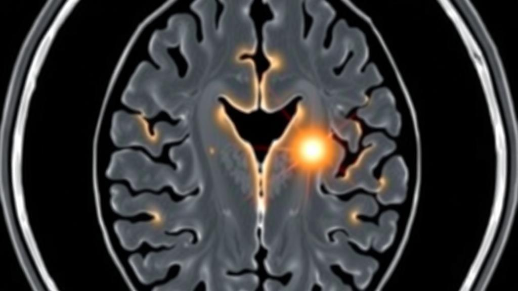
The most clinically significant contribution, however, may be the framework’s transparency layer. The team applied Gradient-weighted Class Activation Mapping, known as Grad-CAM, a technique that exploits the gradients flowing through the final convolutional layers of the network to compute a heatmap of which image regions contributed most strongly to each prediction. When the model flags a scan as glioma, the Grad-CAM overlay highlights the exact area of the brain that drove that conclusion, allowing a radiologist to verify in seconds whether the algorithm is attending to a genuine lesion or, worryingly, to some irrelevant artifact such as a scanner marker or skull edge. This transforms the AI from an oracle issuing unverifiable verdicts into a colleague whose reasoning can be checked.
That transparency was put to the test with expert assessment. A senior medical officer with clinical expertise reviewed representative test cases and evaluated whether the model’s predictions, the Grad-CAM visualizations, and standard clinical judgment aligned. The assessment found strong agreement among all three, supporting the claim that the model’s explanations are not merely visually plausible but clinically meaningful. In the emerging field of explainable AI for healthcare, this human-in-the-loop validation step is what separates a genuine decision-support tool from a demo that looks impressive but collapses under expert scrutiny.
The clinical stakes of this work are considerable. Gliomas, meningiomas, and pituitary tumors each demand different treatment pathways, from surveillance to surgery to radiation planning, and misclassification at the imaging stage can cascade into delayed or inappropriate care. Glioblastoma, the most aggressive glioma, carries a notoriously poor survival rate that improves with earlier detection. An automated system that reliably triages scans, flags suspicious findings, and explains itself to the reviewing physician could shorten the path from initial imaging to definitive diagnosis, particularly in hospitals and regions where subspecialty neuroradiology expertise is scarce. The framework’s millisecond inference time makes it feasible to embed such triage directly into the picture archiving systems that hospitals already use.
The researchers have also made the study unusually reproducible. All three source datasets are publicly available on Kaggle and Figshare, and the merged dataset used in the experiments is itself published, allowing other teams to replicate the benchmark and compare their own architectures against FT_InceptionV3 under the same conditions. The source code is available from the corresponding author on reasonable request. The authors note that the study used anonymized MRI data from public repositories and involved no direct participation of human subjects, and they declare no competing interests. As explainable AI continues its migration from computer science conferences into hospital corridors, this study offers a template for what credible clinical AI research should look like: rigorous benchmarking, honest validation, fast enough for the real world, and, crucially, willing to show its work.
Subject of Research: Explainable deep learning for brain tumor classification from MRI images
Article Title: An explainable fine-tuned deep learning network for precise brain tumor detection from MRI images
Article References: An explainable fine-tuned deep learning network for precise brain tumor detection from MRI images. (n.d.). https://doi.org/10.1007/s11042-026-21919-x
Image Credits: AI Generated
DOI: 10.1007/s11042-026-21919-x
Keywords: brain tumor, deep learning, explainable AI, MRI, InceptionV3, Grad-CAM, transfer learning, convolutional neural network, medical imaging, clinical decision support, cross-validation, data augmentation
Cite Scienmag News
Cassandra Pierce. (October 2, 2026). Explainable AI Spots Brain Tumors on MRI Scans in Milliseconds. Scienmag. https://scienmag.com/explainable-ai-spots-brain-tumors-on-mri-scans-in-milliseconds/
Cassandra Pierce. "Explainable AI Spots Brain Tumors on MRI Scans in Milliseconds." Scienmag, 2 October 2026, https://scienmag.com/explainable-ai-spots-brain-tumors-on-mri-scans-in-milliseconds/. Accessed 2 October 2026.
Cassandra Pierce. "Explainable AI Spots Brain Tumors on MRI Scans in Milliseconds." Scienmag. October 2, 2026. https://scienmag.com/explainable-ai-spots-brain-tumors-on-mri-scans-in-milliseconds/


