


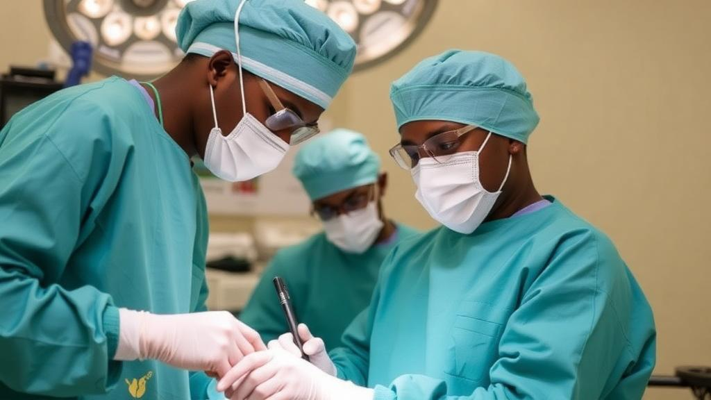
In operating theatres in Nairobi, a team of reconstructive surgeons has demonstrated that one of the most technically demanding procedures in modern medicine—microsurgical free flap reconstruction of the lower limb—can be performed successfully in a health system where resources are scarce and such operations are often considered out of reach. A newly published case series in BMC Plastic and Reconstructive Surgery documents fourteen consecutive lower extremity free flap reconstructions carried out at two tertiary trauma centres, Kenyatta National Hospital and Aga Khan University Hospital, between 2022 and 2025. The results, which include a high rate of flap survival and successful limb salvage in carefully selected patients, challenge a long-standing assumption that advanced microsurgery must remain the preserve of wealthy, well-equipped health systems.
The clinical problem the surgeons confronted is enormous. Lower extremity injuries, particularly those caused by road traffic accidents, are the second leading cause of morbidity and mortality in Kenya, and open fractures with extensive soft tissue loss are among the most difficult injuries in all of trauma surgery. These are dynamic wounds that evolve over hours after the initial insult, and their management demands a multidisciplinary approach involving both orthopaedic and plastic surgeons, rigorous infection control, skeletal stabilization, and ultimately durable soft tissue coverage. When local tissue is insufficient to cover exposed bone, tendons, and hardware, the gold standard is free tissue transfer: harvesting a segment of muscle or skin, along with its artery and vein, from a distant part of the body and reconnecting its blood vessels to vessels at the recipient site under magnification.
Free flap surgery of this kind routinely achieves success rates exceeding 90 per cent in high-income countries, and it has measurably reduced amputation rates for severely injured limbs. Yet the barriers to replicating these outcomes in low- and middle-income countries are formidable. Operating microscopes are expensive to purchase and maintain, microsurgical instruments are costly, and trained microsurgeons are scarce. Many reconstructive units in sub-Saharan Africa lack access to even the basic infrastructure required, and patients frequently receive suboptimal treatment, delayed referrals, or primary amputation as the default option. The Kenyan team set out to show what is achievable when a well-equipped tertiary centre, staffed by experienced microsurgeons, exists within an otherwise resource-limited system.
The series comprised fourteen adult patients, ten male and four female, all of whom underwent lower extremity free flap reconstruction with at least one month of follow-up. Eleven had Gustilo-Anderson type 2-3b open fractures, two had degloving injuries in which skin and underlying tissue were torn away from the limb, and one required reconstruction after removal of a tumour of the hallux, the big toe. Preoperative vascular assessment included computed tomography angiography and handheld Doppler ultrasonography—diagnostic resources that are rarely available in comparable settings and that proved critical for mapping recipient vessels before the operating room. All procedures were performed by a consultant microsurgeon at a Level 1 trauma facility, under general anaesthesia, with wound debridement conducted under tourniquet control.
The technical details of the operations reveal the precision the procedure demands. Fasciocutaneous free flaps, which carry skin and its underlying fascia, were used in nine patients, while muscle flaps were used in five. Recipient vessels were selected according to wound configuration, local anatomy, and the mechanism of injury, with surgeons carefully examining the vessel wall, the extent of injury, and the quality of the adventitia before committing to an anastomosis. Crucially, all arterial connections were performed proximal to the zone of injury, following the principle that vessels damaged by trauma provide unreliable inflow. Thirteen cases used end-to-end anastomoses to the anterior tibial artery, the dorsalis pedis artery, or the superior medial genicular artery, while one case used an end-to-side connection to the posterior tibial artery. Where a surgical microscope was unavailable, surgeons achieved comparable results using loupe magnification in five cases.
The case series includes vivid individual examples that illustrate the range of reconstructive options. One middle-aged woman suffered a left ankle degloving injury, a distal tibia fracture, and complex bilateral midfoot fracture-dislocations in a road traffic accident. After initial washout, external fixation, and vacuum-assisted closure dressings, the team performed delayed definitive closure using a free gracilis muscle flap—the posterior-most adductor muscle of the thigh, supplied by the medial circumflex femoral artery with a pedicle roughly seven centimetres long—combined with a skin graft and internal fixation of the fractures. A two-team approach allowed simultaneous harvest and recipient-site preparation, shortening operative time and reducing donor-site morbidity. The flap integrated fully, with satisfactory functional and aesthetic outcomes at follow-up.
Another patient, a soldier with blast injuries to the right foot, presented with segmented fractures of the metatarsals and midfoot bones, bony defects at the second and third metatarsals, and loss of Lisfranc alignment. After debridement, K-wire stabilization, and temporary vacuum dressings, the surgeons planned a latissimus dorsi free muscle flap based on the thoracodorsal artery, whose single dominant pedicle can reach twelve centimetres in length and 2.5 millimetres in diameter. The flap was designed with a lazy-S skin paddle oriented along the pedicle, the serratus branch was preserved for venous anastomosis, and intraoperative imaging confirmed the muscle flap in place alongside antibiotic bone cement. A third patient, who developed chronic osteomyelitis after a high-energy motor vehicle collision, underwent a first-stage Masquelet procedure with a cement spacer and soft tissue coverage using a parascapular free flap harvested through the triangular space of the back. A fourth, whose foot was crushed in an industrial accident, received a free anterolateral thigh flap over a transmetatarsal amputation stump, with a total ischemic time of just one hour.
Postoperative care followed a rigorous, standardized protocol. Patients were admitted to the critical care unit for forty-eight hours, where flaps were assessed hourly by an attending consultant and a senior registrar, with checks of colour, capillary refill, temperature, and Doppler signals to detect early vascular compromise. Venous thromboembolism prophylaxis consisted of enoxaparin 40 milligrams subcutaneously once daily, alongside aspirin 75 milligrams, and limbs were elevated and immobilized in splints. Mobilization began on postoperative day five with individualized protocols and compression bandaging. This intensity of monitoring is itself notable: in many low-resource settings, patients are not routinely admitted to critical care, and flaps are monitored on general wards using clinical parameters alone, including pinprick testing for bleeding quality.
Outcomes were strong but not flawless. Twelve of the fourteen flaps survived without complication. Two flaps failed—one attributed to suspected underlying peripheral arterial disease, detected intraoperatively as weak dorsalis pedis flow and dark venous bleeding despite an intact pedicle, and the other to multifactorial causes—both necessitating debridement and skin graft reconstruction. Definitive management ranged from ten to fifty-six days, with an average hospital stay of two to three weeks. All patients completed at least one month of follow-up without delayed flap failures. The authors acknowledge limitations, including the absence of diagnostic angiograms for preoperative detection of peripheral arterial disease, the inability to formally assess functional outcomes, and loss of some patients to long-term follow-up.
The broader significance of the study lies in what it suggests about global surgical equity. The flap survival rates achieved in Nairobi matched those reported in high-income countries, demonstrating that successful microsurgical reconstruction is possible in resource-limited settings when expertise and infrastructure converge in a well-equipped tertiary centre. The authors argue that expanding fellowship opportunities for reconstructive surgeons in low- and middle-income countries is essential, and that surgeons returning from such training can practise microsurgical techniques using basic materials and shared local resources. They also call for structured retrospective and prospective observational studies to build the evidence base further. For the millions of patients worldwide who suffer devastating limb injuries each year, the message is clear: the decision between salvage and amputation should depend on clinical factors and access to skilled teams—not on geography or the wealth of the health system a patient happens to be born into.
Subject of Research: Lower extremity free flap reconstruction for traumatic limb injuries in a resource-limited health system
Article Title: Lower extremity free flap reconstructions in a tertiary trauma centre within a resource-limited health system: a case series
Article References: Otieno, D. O., Okello, A. W., Gathura, E. W., Gebremariyam, Z. T., Nang’ole, F. W., Sulemanji, D. S., & Wabwire, B. (2026). Lower extremity free flap reconstructions in a tertiary trauma centre within a resource-limited health system: a case series. BMC Plastic and Reconstructive Surgery, 2(1), Article 3. https://doi.org/10.1186/s44452-026-00015-x
Image Credits: AI Generated
DOI: 10.1186/s44452-026-00015-x
Keywords: free flap reconstruction, microsurgery, lower extremity trauma, limb salvage, resource-limited settings, Kenya, open fractures, reconstructive surgery, trauma surgery, fasciocutaneous flaps, muscle flaps, global surgery
Cite Scienmag News
Ophelia Keating. (October 1, 2026). Surgeons in Kenya Prove Complex Limb-Saving Microsurgery Works in Resource-Limited Settings. Scienmag. https://scienmag.com/surgeons-in-kenya-prove-complex-limb-saving-microsurgery-works-in-resource-limited-settings/
Ophelia Keating. "Surgeons in Kenya Prove Complex Limb-Saving Microsurgery Works in Resource-Limited Settings." Scienmag, 1 October 2026, https://scienmag.com/surgeons-in-kenya-prove-complex-limb-saving-microsurgery-works-in-resource-limited-settings/. Accessed 1 October 2026.
Ophelia Keating. "Surgeons in Kenya Prove Complex Limb-Saving Microsurgery Works in Resource-Limited Settings." Scienmag. October 1, 2026. https://scienmag.com/surgeons-in-kenya-prove-complex-limb-saving-microsurgery-works-in-resource-limited-settings/


