


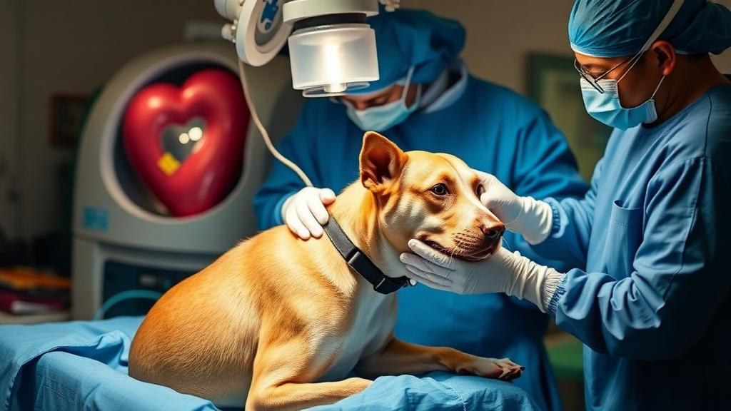
In a striking example of how advanced human cardiac surgery techniques are finding their way into veterinary medicine, a surgical team has reported the successful palliative debulking of a heart-base tumour that had invaded the right atrium of a French Bulldog, using partial cardiopulmonary bypass. The case, published in Veterinary Medicine and Science, describes what the authors believe is the first English-language report of a canine aortic body tumour extending into the right atrial lumen being surgically debulked with the support of a heart-lung machine. The operation did not cure the dog, but it bought him months of comfortable life by relieving a life-threatening mechanical obstruction that medication alone could not touch.
Aortic body tumours, also called chemodectomas, are slow-growing neoplasms that arise from chemoreceptor cells located near the base of the heart, close to the aorta. In dogs they are typically locally expansive masses that cause trouble by compressing adjacent cardiovascular structures rather than by invading the heart chambers themselves. Intracavitary or intracardiac involvement is genuinely uncommon, and previous reports of intracardiac extension did not describe tumour debulking under cardiopulmonary bypass. Cardiopulmonary bypass, or CPB, has been used in dogs for selected intracardiac procedures, including removal of a left atrial paraganglioma and a tricuspid valve myxoma, but a partial-bypass palliative debulking of a heart-base tumour protruding into the right atrium had not previously been documented.
The patient was a seven-year-old, 15.2-kilogram castrated male French Bulldog referred for lethargy, poor appetite, exercise intolerance and abdominal distension of roughly two weeks’ duration. Ultrasound had revealed free fluid in the abdomen, and echocardiography showed severe right-sided cardiac enlargement together with a mass measuring 3.2 by 3.1 centimetres occupying most of the right atrial lumen. The mass was heterogeneously echogenic with an irregular surface, appeared to originate from the heart base adjacent to the aorta, and was associated with significant tricuspid regurgitation and paradoxical septal motion. Abdominal imaging showed hepatic venous congestion and a large volume of ascites, and roughly 600 millilitres of fluid was drained to relieve the distension. The fluid was classified as a modified transudate, consistent with back-pressure from an obstructed right atrium rather than primary abdominal disease.
The clinical picture was one of mechanical obstruction of venous return. Blood attempting to flow from the caudal vena cava into the right atrium was blocked by the intracavitary mass, raising right atrial pressure, congesting the hepatic veins and driving fluid out into the peritoneal cavity. Medical management with pimobendan, furosemide and low-molecular-weight heparin failed to resolve the ascites or improve the dog’s activity and appetite, which strengthened the suspicion that the problem was structural rather than functional. Differential diagnoses included neoplasia, thrombus and tumour-associated thrombus, but antithrombotic therapy produced no reduction in the mass. Preoperative biopsy was judged too hazardous because the mass sat within the right atrium adjacent to critical structures, so the team proceeded without a definitive tissue diagnosis, informing the owner that the operation would be palliative rather than curative.
The surgical plan was deliberately unconventional. After premedication with midazolam, fentanyl and cefazolin, anaesthesia was induced with propofol and maintained with sevoflurane. The right jugular vein and right carotid artery were exposed for cannulation: a 14-Fr venous drainage cannula was advanced through the cranial vena cava to near the right atrial junction, and an 8-Fr arterial cannula was placed in the carotid artery. A right fifth intercostal thoracotomy exposed the heart. Notably, the team attempted additional venous cannulation from the right atrium toward the caudal vena cava but could not advance the cannula because the mass was in the way. Partial CPB was therefore run on the cervical cannulas alone, with heparin achieving activated clotting times above 400 seconds and later around 1000 seconds. Pump flow was set at 60 to 80 millilitres per kilogram per minute, and the dog was cooled to a lowest recorded oesophageal temperature of 28 degrees Celsius.
The authors made two decisions that reveal the improvisational nature of the procedure. First, the azygos vein was deliberately left unoccluded. Although temporarily occluding it would have reduced venous return into the operative field, venous return from the caudal body was already compromised by the mass, so preserving collateral drainage through the azygos system was considered preferable even at the cost of some surgical visibility. Second, after debulking improved caudal vena caval return, the team chose not to pursue additional cannulation, instead managing venous blood with two suction lines whose contents were returned to the extracorporeal circuit. Perfusion was judged adequate on the basis of arterial pressure, serial blood gas analyses and lactate concentrations, which remained between 1.0 and 2.5 mmol/L throughout.
Opening the right atrium revealed a mass firmly adherent to the septal aspect of the chamber, with marked induration suggesting neoplastic tissue. The surgeons used Babcock forceps to gently stabilise the intracavitary component and removed tumour primarily by blunt dissection with careful sharp dissection near the attachment site. No distinct dissection plane existed in the firmly adherent portion, so complete separation was not attempted. Residual tumour was intentionally left in place wherever further dissection risked perforation, uncontrollable haemorrhage or injury to critical structures such as the coronary sinus or atrioventricular junction. The atriotomy was closed in two layers with 6-0 polypropylene. Approximately ten minutes after atriotomy, ventricular fibrillation occurred; defibrillation after closure restored sinus rhythm. Total anaesthesia time was 410 minutes, surgical time 350 minutes, CPB time 140 minutes and intracardiac manipulation time just 40 minutes.
The haemodynamic payoff was immediate and measurable. Intermittently recorded central venous pressure fell from approximately 19 mmHg before debulking to approximately 5 mmHg afterwards, while systemic arterial pressure actually rose. Postoperatively, the mucous membrane pallor resolved, the ascites disappeared, and appetite and activity returned. Pimobendan and furosemide were discontinued, clopidogrel was prescribed for about a month, and follow-up echocardiography showed only minimal residual intracardiac tissue, reduced right-sided enlargement and no detectable tricuspid regurgitation. The dog was discharged on day 14. Histopathology of the resected tissue suggested a neuroendocrine neoplasm, and immunohistochemistry positive for synaptophysin and chromogranin A supported the diagnosis. Toceranib, a tyrosine kinase inhibitor with reported benefit in canine chemodectomas, was started on day 21.
The longer arc of the case tempers the enthusiasm. On day 360, a recurrent right atrial mass measuring 2.0 by 1.9 centimetres was documented, although without signs of right-sided heart failure. On day 390 the dog developed pancreatitis with biliary obstruction and died of multiple organ failure. Necropsy confirmed an aortic body tumour infiltrating from the endocardial surface into the right atrial myocardium, with pulmonary metastases and tumour emboli in the left caudal lung lobe. The authors are careful to note that they cannot determine whether the pulmonary spread represented spontaneous disease progression or was partly procedure-associated, because suctioned blood was returned to the circuit and standard filters cannot reliably remove individual tumour cells. They also acknowledge that the contribution of surgery, toceranib and the tumour’s naturally slow growth to the dog’s ten-month survival cannot be disentangled.
The team is equally clear that this should not be read as a new standard of care. Preoperative CT staging was not performed, central venous pressure was not monitored during bypass, and no strict numerical abort criterion was set, all acknowledged limitations. Total venous inflow occlusion was considered as a simpler alternative but rejected because the mass required careful stepwise debulking rather than rapid excision, and the 40-minute intracardiac manipulation exceeded the short window that technique allows. The authors frame the operation as an exceptional, high-risk rescue intervention justified only when severe intracardiac obstruction is the dominant life-limiting problem, medical and radiation options cannot provide rapid relief, and advanced cardiac surgical and perfusion support are available. Within those narrow boundaries, the case demonstrates that partial cardiopulmonary bypass can deliver real, if temporary, palliative benefit in veterinary patients facing obstruction that would otherwise be fatal.
Subject of Research: Palliative surgical debulking of a canine aortic body tumour with right atrial invasion under partial cardiopulmonary bypass
Article Title: Palliative Debulking of an Aortic Body Tumour With Right Atrial Invasion in a Dog Under Partial Cardiopulmonary Bypass
Article References: Suzuki, S., Tanaka, S., Kanno, N., Yogo, T., Harada, Y., Michishita, M., & Hara, Y. (2026). Palliative Debulking of an Aortic Body Tumour With Right Atrial Invasion in a Dog Under Partial Cardiopulmonary Bypass. Veterinary Medicine and Science, 12(5), Article e71244. https://doi.org/10.1002/vms3.71244
Image Credits: AI Generated
DOI: 10.1002/vms3.71244
Keywords: aortic body tumour, chemodectoma, cardiopulmonary bypass, right atrium, veterinary surgery, French Bulldog, cardiac tumour, ascites, toceranib, palliative care, echocardiography, neuroendocrine tumour
Cite Scienmag News
William Thompson. (October 1, 2026). Surgeons Use Heart-Lung Machine to Debulk Rare Cardiac Tumour in a Dog. Scienmag. https://scienmag.com/surgeons-use-heart-lung-machine-to-debulk-rare-cardiac-tumour-in-a-dog/
William Thompson. "Surgeons Use Heart-Lung Machine to Debulk Rare Cardiac Tumour in a Dog." Scienmag, 1 October 2026, https://scienmag.com/surgeons-use-heart-lung-machine-to-debulk-rare-cardiac-tumour-in-a-dog/. Accessed 1 October 2026.
William Thompson. "Surgeons Use Heart-Lung Machine to Debulk Rare Cardiac Tumour in a Dog." Scienmag. October 1, 2026. https://scienmag.com/surgeons-use-heart-lung-machine-to-debulk-rare-cardiac-tumour-in-a-dog/


