


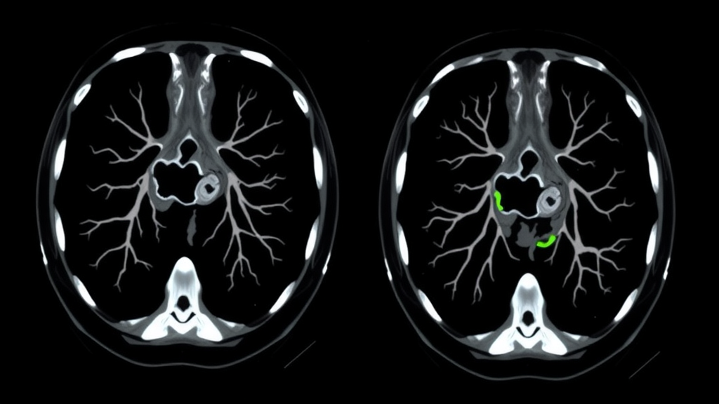
A routine workup for a stubborn, low-grade fever and purulent sputum in a 41-year-old woman has led clinicians to one of the rarest presentations of a congenital lung anomaly ever described, according to a case report published in Respirology Case Reports. The woman, referred for surgical treatment of suspected pulmonary sequestration, was ultimately found to have congenital bronchial atresia confined to Segment 7 of her right lower lobe — a developmental defect so uncommon in this location that it can convincingly impersonate a completely different malformation on standard imaging.
Congenital bronchial atresia, or CBA, is a rare developmental anomaly in which a lobar, segmental or subsegmental bronchus is focally interrupted while the distal bronchial tree remains intact. Because the airway is blocked proximally, mucus accumulates downstream, forming a mucus-filled cavity known as a bronchocele or mucocele. At the same time, air finds its way into the affected tissue through collateral ventilation channels, causing the trapped lung segment to become hyperinflated. The pairing of a bronchocele with distal hyperinflation is considered the radiological fingerprint of the condition, and in most cases computed tomography can nail the diagnosis by showing the abrupt bronchial interruption, the mucus-filled structure and the adjacent hyperlucent, emphysematous lung.
But the clinical picture becomes murkier when the anomaly sits in the lower lobes. Pulmonary sequestration, a key differential diagnosis for lower-lobe congenital lung lesions, is defined by the complete absence of normal bronchial communication and the presence of an aberrant systemic arterial supply, typically arising from the aorta. Lower-lobe CBA can also present with recurrent infection and cystic or cavitary changes, meaning the two conditions can look strikingly similar when inflammation blurs the anatomical details. That is precisely what happened in this case, and the report underscores why a systematic evaluation of both bronchial continuity and vascular supply is essential before any diagnosis of a congenital lower-lobe lesion is finalized.
The patient’s story began with annual episodes of respiratory infection, each temporarily quieted by oral antibiotics. Chest radiography revealed an abnormal shadow in the right lung field, and CT imaging demonstrated a cavitary lesion with internal fluid retention in the right lower lobe, surrounded by ground-glass opacity suggestive of active inflammation. After a course of antibiotic therapy settled the inflammation, she was referred for surgical management, still carrying the presumptive diagnosis of pulmonary sequestration.
Repeat computed tomography at the treating institution changed the picture dramatically. The scans revealed segmental emphysematous change localized mainly to Segment 7 of the right lower lobe, alongside the cavitary lesion with internal fluid retention and faint residual ground-glass opacity. Critically, the imaging identified an interruption of the right B7 bronchus — the segmental airway serving Segment 7. Just as importantly, no aberrant systemic arterial supply from the aorta was observed anywhere in the region. Instead, the pulmonary artery showed a normal branching pattern, with a normally distributed segmental artery, designated A7, supplying the lesion. Together, these findings pointed decisively away from pulmonary sequestration and toward congenital bronchial atresia, with the cavitary lesion reinterpreted as a bronchocele.
Three-dimensional CT reconstruction sealed the diagnosis. The technique clearly demonstrated the interruption of the B7 bronchus and the normal distribution of the A7 segmental artery, while the pulmonary venous anatomy was confirmed to be unremarkable. This imaging approach, the report notes, is particularly valuable when sublobar resection is being considered, because precise anatomical understanding of the bronchovascular architecture is required for safe and appropriate surgery. In this patient, however, the geometry of the disease made lung-sparing surgery impractical: although the lesion was centered in Segment 7, the hyperinflation extended close to the basal bronchial bifurcation, and inflammatory changes on earlier scans had involved the adjacent segments. Complete resection by segmentectomy was therefore judged too difficult, and the surgical team opted for a thoracoscopic right lower lobectomy.
The operation itself went smoothly. Pulmonary function testing before surgery had been within normal limits, and laboratory findings showed only mild inflammatory change. Intraoperative bronchoscopy confirmed the imaging findings, demonstrating no identifiable orifice of the B7 bronchus at the level of the right intermediate bronchus, while the orifices of B6 and the remaining basal bronchi, B8 through B10, were clearly visible. The procedure took 123 minutes with minimal blood loss, the postoperative course was uneventful, and the patient was discharged on the eighth day after surgery.
Histopathological examination of the resected tissue provided the final confirmation. The cavitary lesion was composed of bronchial structures filled with retained mucus and showed no communication with the central airway — exactly the architecture expected of a bronchocele associated with congenital bronchial atresia. Based on the combined radiological, bronchoscopic and histopathological findings, the team established the final diagnosis of CBA, closing a diagnostic journey that had begun with a completely different suspected condition.
The case carries several important lessons for clinicians and radiologists. Although the combination of a bronchocele and distal hyperinflation is characteristic of CBA, a lower-lobe location, recurrent infection and a cavitary appearance can cause the anomaly to radiologically resemble pulmonary sequestration. The distinction matters enormously: pulmonary sequestration accounts for approximately 0.15 percent to 6.4 percent of congenital pulmonary malformations and is defined by its aberrant systemic blood supply, whereas CBA preserves normal pulmonary arterial supply despite its proximal bronchial interruption. In CBA, the bronchus is interrupted proximally while the distal airway is preserved and filled with mucus; in sequestration, there is no normal bronchial communication at all, and the tissue is fed by a systemic artery, typically from the aorta.
Rarity compounds the diagnostic difficulty. CBA is less frequently encountered in clinical practice than sequestration and is most commonly detected incidentally, with a strong predilection for the upper lobes — particularly the left upper lobe. Lower-lobe involvement is uncommon in adult radiologic series, and segmental bronchial atresia confined to a single basal segment such as S7 is extremely rare. In such cases, recurrent infection and cystic or cavitary changes can closely resemble intralobar pulmonary sequestration, making accurate differentiation genuinely challenging even for experienced teams. The authors emphasize that systematic evaluation of both bronchial continuity and vascular supply is the key to getting it right, and that three-dimensional CT reconstruction may prove useful not only for diagnosis but also for guiding surgical decision-making, from choosing between segmentectomy and lobectomy to mapping the precise anatomy the surgeon will encounter.
Subject of Research: Congenital bronchial atresia of the right lower lobe mimicking pulmonary sequestration
Article Title: Congenital Bronchial Atresia Mimicking Pulmonary Sequestration in the Lower Lobe: A Case Report
Article References: Mitsumata, S., Midorikawa, K., Wakamatsu, K., Kaneda, S., Futamata, T., Iwanaka, T., Ueda, Y., Miyahara, S., Masuda, Y., Nakashima, H., & Sato, T. (2026). Congenital Bronchial Atresia Mimicking Pulmonary Sequestration in the Lower Lobe: A Case Report. Respirology Case Reports, 14(9), Article e70712. https://doi.org/10.1002/rcr2.70712
Image Credits: AI Generated
DOI: 10.1002/rcr2.70712
Keywords: congenital bronchial atresia, pulmonary sequestration, bronchocele, three-dimensional CT reconstruction, thoracoscopic lobectomy, segment 7 right lower lobe, recurrent lung infection, bronchial interruption, segmental emphysema, congenital pulmonary malformation, surgical planning, case report
Cite Scienmag News
Ophelia Keating. (September 23, 2026). Rare Congenital Lung Anomaly Masquerades as a Lookalike Condition on CT Scans. Scienmag. https://scienmag.com/rare-congenital-lung-anomaly-masquerades-as-a-lookalike-condition-on-ct-scans/
Ophelia Keating. "Rare Congenital Lung Anomaly Masquerades as a Lookalike Condition on CT Scans." Scienmag, 23 September 2026, https://scienmag.com/rare-congenital-lung-anomaly-masquerades-as-a-lookalike-condition-on-ct-scans/. Accessed 23 September 2026.
Ophelia Keating. "Rare Congenital Lung Anomaly Masquerades as a Lookalike Condition on CT Scans." Scienmag. September 23, 2026. https://scienmag.com/rare-congenital-lung-anomaly-masquerades-as-a-lookalike-condition-on-ct-scans/


