


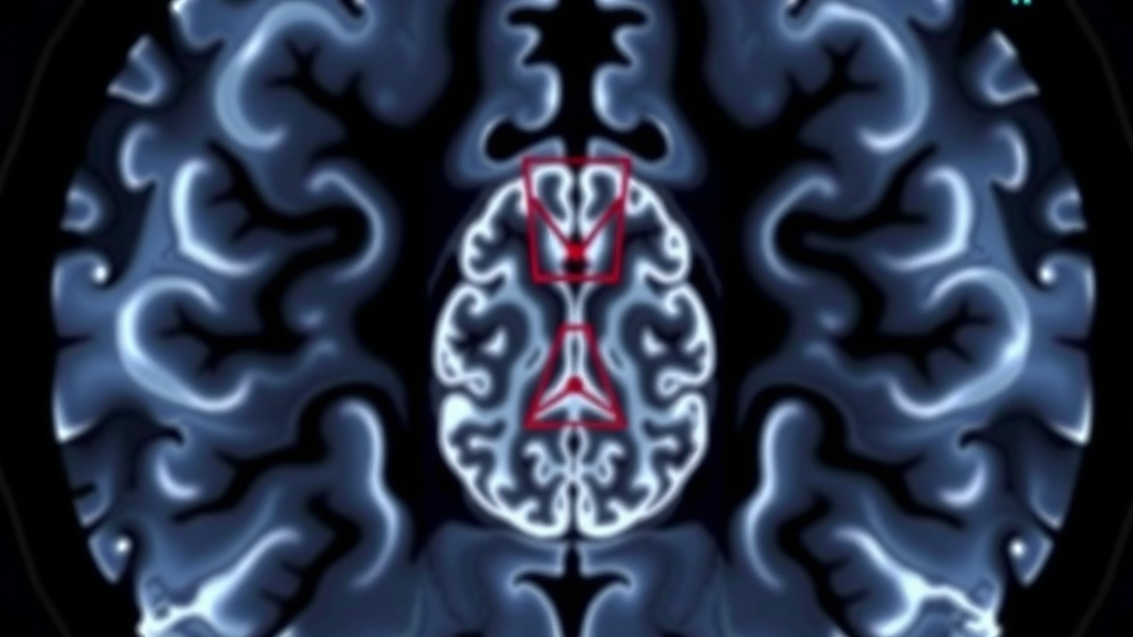
A faint whisper of contrast enhancement on a diagnostic MRI scan may be the single most powerful warning sign that a seemingly indolent brain tumor is poised to turn malignant, according to a new study that also delivers the first statistically rigorous estimates of when that transformation actually occurs. The research, published in the Journal of Neuro-Oncology by a team at Kepler University Hospital and Johannes Kepler University Linz, followed 155 patients with WHO grade 2 or 3 diffuse gliomas — often called lower-grade gliomas — using a battery of four complementary survival models and, for the first time in this field, treating malignant transformation as an interval-censored event whose true timing lies somewhere between two consecutive scans.
Lower-grade gliomas occupy an uneasy middle ground in neuro-oncology. They can smolder quietly for years, sometimes over a decade, allowing patients to work, drive and live largely normal lives under watchful surveillance. Yet nearly half of the patients in the new cohort — 77 of 155 — eventually underwent malignant transformation, the biological inflection point in which a slow-growing tumor acquires the aggressive behavior of a high-grade glioma and drives prognosis, adjuvant-therapy decisions and eligibility for clinical trials. Predicting which tumors will take that turn, and when, has remained one of the field’s most stubborn uncertainties, largely because transformation can only be pinned down retrospectively, at the moment a confirmatory MRI or a repeat biopsy reveals it.
The Austrian-led team attacked this problem from several angles simultaneously. Every patient had histologically confirmed adult-type diffuse glioma, diagnostic-quality multiparametric MRI including pre- and post-contrast T1, T2 and FLAIR sequences, and at least one follow-up scan. The time origin for each patient was the first MRI showing the glioma — a deliberately chosen anchor, since 18 patients in the cohort were managed with a wait-and-see strategy for more than a year, with one waiting 17.3 years before histological confirmation, contributing their untreated natural history to the analysis. Because malignant transformation is only ascertainable between examinations, the researchers bracketed each event within an interval bounded by the last documentation of low-grade status and the first examination showing transformation, rather than falsely assigning the event to a single scan date.
That methodological choice mattered enormously. When the team compared conventional dating — which treats the confirmatory scan as the moment of transformation — with proper interval-censored analysis, the conventional approach overstated the hazard of transformation roughly four-fold. The finding exposes a systematic bias running through much of the existing literature, where standard right-censored Cox models equate radiological detection with biological occurrence, an error whose magnitude grows with the length of the surveillance interval. To ensure their conclusions were not artifacts of any single statistical specification, the investigators cross-checked the primary Bayesian interval-censored Weibull model, fitted with a regularized horseshoe prior across 102 candidate predictors, against three frequentist alternatives: a Fine-Gray subdistribution-hazards model accounting for the competing risk of death without transformation, a Cox counting-process model handling surgery as a time-varying covariate with 20-fold multiple imputation, and a native interval-censored Fine-Gray model estimated by sieve maximum likelihood. All four agreed on the direction and significance of the central result.
And that central result was striking. Contrast enhancement visible on the diagnostic scan — even the subtle, sub-threshold kind that does not itself meet radiological criteria for malignancy — was by far the strongest predictor of subsequent malignant transformation, dwarfing both tumor volume and a co-analyzed panel of 99 quantitative radiomic features. In the full cohort, the Bayesian hazard ratio for enhancement reached 22.1, with a 95 percent credible interval of 10.5 to 53.2. Within IDH-mutant disease the association approached separation: all 22 enhancing tumors eventually transformed, compared with only 30 percent of non-enhancing ones, a pattern so extreme that the statisticians had to deploy Firth penalized-likelihood and exact conditional methods to obtain interpretable lower bounds. The same held true across every molecular entity represented — IDH-mutant astrocytomas, 1p/19q-codeleted oligodendrogliomas and IDH-wildtype tumors — with every single enhancing tumor in each subgroup ultimately transforming.
Critically, the team recognized that much of this dramatic unadjusted effect reflects tumors that were already transforming at or near diagnosis, so they performed a prespecified series of landmark analyses that condition on surviving transformation-free for three, six or twelve months and restart the clock there. Even under this stricter framing, a five- to seven-fold elevation in hazard persisted at every landmark: 7.41 at three months, 5.65 at six months and 6.33 at twelve months. The authors note that an unmeasured confounder would need an implausibly strong association with both enhancement and transformation — a risk ratio of at least 5.8 with each — to explain away the six-month estimate. Blinded re-reading of baseline scans by two independent reviewers achieved perfect agreement on enhancement status, and sensitivity analyses excluding patients who would meet the 2021 WHO grade 4 criteria left the effect essentially unchanged.
Perhaps the most surprising discovery concerned what happened after surgery. Because the exposure and the endpoint are both read from post-contrast imaging, one might suspect the enhancement signal simply marks a focus of high-grade disease that a resection removes. The data say otherwise. In a model of time from first surgery restricted to the 128 patients who had not transformed beforehand, baseline enhancement predicted post-surgical malignant transformation with a hazard ratio of 4.28 — and the effect not only persisted within gross-totally resected tumors, where any enhancing tissue present at baseline should have been removed, but strengthened under multivariable adjustment, rising to 7.58 when grade, IDH status and extent of resection were accounted for. Seven of eight enhancing tumors resected gross-totally still transformed, versus only 15 of 60 non-enhancing ones. A signal that outlives the very tissue generating it, the authors argue, points to a property of the tumor as a whole — an intrinsic biology — rather than a discrete focus awaiting excision.
Surgery itself emerged as the only modifiable determinant in the study. Gross-total resection was associated with a roughly two-thirds reduction in the post-surgical hazard of transformation compared with biopsy (hazard ratio 0.32), a benefit also seen within IDH-mutant disease, while subtotal resection showed no significant advantage over biopsy. The finding reinforces the current standard of early onco-functional resection in diffuse glioma, though the authors are careful to note that the timing of surgery in their retrospective cohort was confounded by indication — surgeons often operated precisely because early signs of transformation appeared — and cannot be interpreted causally. What they do propose is that baseline enhancement, currently discarded once histology becomes available, should be carried forward into postoperative risk estimation, where it independently refines prognostication alongside grade, molecular status and resection extent.
The study also delivered a sobering verdict on radiomics. Despite extracting 428 features from co-registered image volumes, reduced to 99 candidates by correlation filtering, not a single quantitative feature survived Bayesian horseshoe shrinkage or native interval-censored analysis. One texture feature retained significance only under the biased midpoint-imputation framework, collapsing to null under proper censoring — a conditional null the authors frame honestly, acknowledging that 77 events cannot credibly support 99 candidate biomarkers, and calling for radiomics discovery to be re-scoped toward predefined panels or much larger cohorts. With a bootstrap-corrected concordance of 0.83 but imperfect absolute-risk calibration, the team explicitly declines to offer their model for individual prediction. Instead, they position the work as a recalibration of the field’s foundations: contrast enhancement on the diagnostic scan deserves a central place in pre- and postoperative risk stratification, gross-total resection materially changes the transformation hazard, and any future estimate of when a lower-grade glioma will turn malignant should respect the interval-censored reality of how that event is actually observed.
Subject of Research: Predictors and timing of malignant transformation in lower-grade glioma using interval-censored survival analysis
Article Title: Malignant transformation of lower-grade glioma: contrast enhancement, extent of resection, and the natural history under interval-censored analysis
Article References: Malignant transformation of lower-grade glioma: contrast enhancement, extent of resection, and the natural history under interval-censored analysis. (n.d.). https://doi.org/10.1007/s11060-026-05803-0
Image Credits: AI Generated
DOI: 10.1007/s11060-026-05803-0
Keywords: lower-grade glioma, malignant transformation, contrast enhancement, MRI, extent of resection, gross-total resection, interval censoring, Bayesian analysis, radiomics, IDH status, WHO classification, brain tumor
Cite Scienmag News
Nathaniel Bowman. (September 22, 2026). One MRI Sign Predicts Which Slow-Growing Brain Tumors Will Turn Deadly. Scienmag. https://scienmag.com/one-mri-sign-predicts-which-slow-growing-brain-tumors-will-turn-deadly/
Nathaniel Bowman. "One MRI Sign Predicts Which Slow-Growing Brain Tumors Will Turn Deadly." Scienmag, 22 September 2026, https://scienmag.com/one-mri-sign-predicts-which-slow-growing-brain-tumors-will-turn-deadly/. Accessed 22 September 2026.
Nathaniel Bowman. "One MRI Sign Predicts Which Slow-Growing Brain Tumors Will Turn Deadly." Scienmag. September 22, 2026. https://scienmag.com/one-mri-sign-predicts-which-slow-growing-brain-tumors-will-turn-deadly/


