


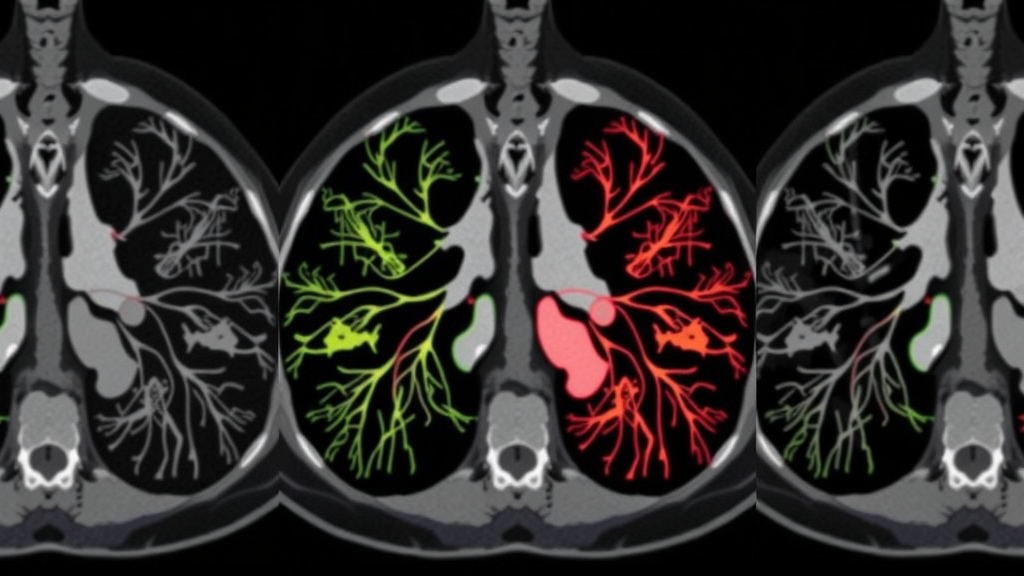
Lung cancer remains one of the deadliest malignancies in the world, and the single biggest reason for its devastating toll is simple: it is usually found too late. By the time symptoms drive a patient to the clinic, the disease has often spread beyond the lung, and the window for curative treatment has narrowed dramatically. Even when patients do undergo screening, distinguishing between the different histological subtypes of lung cancer from computed tomography (CT) images is a subtle, demanding task that challenges even experienced radiologists. Now, a team of researchers from institutions across India has developed a hybrid artificial intelligence model that could make that task faster, more accurate, and more consistent, achieving a testing accuracy of 92 percent in identifying lung cancer and its major subtypes directly from CT scans.
The new study, published in the journal Multimedia Tools and Applications, describes a deep learning architecture that combines two of the most powerful ideas in modern computer vision: convolutional neural networks (CNNs) and transformers. Led by Neha Thakur and colleagues at the Central University of Himachal Pradesh, together with collaborators at VIT Bhopal University and the Central University of Jammu, the work addresses a well-known weakness in the existing literature. While countless studies have applied deep learning to lung cancer imaging, most have simply benchmarked standard architectures without confronting the harder questions of how well a model generalizes to unseen data, whether its features are clinically meaningful, and whether its predictions can be trusted in a hospital setting. Many of those earlier models, the researchers note, reported accuracies below 85 percent, a figure that falls short of what clinicians would consider reliable.
The core innovation of the new framework lies in how it fuses local and global information. Convolutional neural networks are exceptionally good at detecting fine-grained local patterns, such as the texture of a tumor boundary, the density of a nodule, or the spiculated edges that often signal malignancy. Transformers, by contrast, excel at capturing long-range dependencies: they can weigh how a shadow in the upper lobe of one lung relates to structural changes elsewhere in the same scan. Standalone CNNs tend to miss these broader contextual relationships, while pure transformers can struggle with the fine texture detail that matters for distinguishing subtly different tissue types. The hybrid CNN-Transformer model is designed to have the best of both worlds, using convolutional layers for fine-grained texture analysis while attention-based mechanisms capture long-range relationships across regions of lung tissue.
To test the idea, the researchers assembled a benchmark lung cancer CT dataset containing four classes: adenocarcinoma, large cell carcinoma, squamous cell carcinoma, and normal lung tissue. These categories are not arbitrary labels; they correspond to biologically distinct diseases with different treatment pathways and prognoses. Adenocarcinoma is the most common form of non-small cell lung cancer and often arises in the outer regions of the lung, while squamous cell carcinoma tends to develop in the central airways and is strongly associated with smoking history. Large cell carcinoma is a less common but particularly aggressive variant. Getting the subtype right matters enormously, because targeted therapies, surgical decisions, and chemotherapy regimens all depend on an accurate histological diagnosis.
Before any learning took place, the team invested heavily in data preparation. Extensive pre-processing and data normalization were applied to the CT images, and the model’s hyperparameters were carefully tuned to maximize robustness and reduce the risk of overfitting, the common failure mode in which a network memorizes its training examples rather than learning genuinely transferable patterns. This methodical pipeline reflects a growing recognition in the medical AI community that architectural novelty alone is not enough; the quality and consistency of the input data and the discipline of the training regime often determine whether a model performs well in practice or merely in a paper.
The benchmarking itself was unusually thorough. The hybrid CNN-Transformer was compared not only against a conventional CNN but also against recurrent architectures including LSTM, CNN-LSTM, and BiLSTM, as well as the leading transformer-based vision models: Vision Transformer (ViT), Data-efficient Image Transformer (DeiT), and Swin Transformer. Across all comparisons, the hybrid architecture emerged as the strongest performer, achieving 92 percent testing accuracy, a weighted precision of 93 percent, recall of 92 percent, and an F1-score of 92 percent. These are balanced scores across all four classes, which is significant: it means the model is not simply succeeding on easy categories while collapsing on hard ones, a flaw that has undermined many earlier multi-class medical imaging systems.
The detailed error analysis reinforced that picture. Confusion matrices, which record exactly where the model’s predictions diverged from the true labels, and precision-recall analyses both showed improved class separability, with particularly strong gains on the clinically significant subtypes of squamous cell carcinoma and adenocarcinoma. In other words, the model was at its most reliable precisely where mistakes carry the highest clinical cost. The researchers interpret this as evidence that the combined architecture produces a more balanced and discriminative internal representation of the imaging data than either standalone models or the other hybrids they tested, allowing it to tease apart tissue patterns that other networks blur together.
The implications for clinical practice could be substantial. A decision-support system of this kind would not replace the radiologist or pathologist, but it could act as a tireless second reader, flagging suspicious scans for priority review, suggesting a likely subtype to guide the ordering of confirmatory biopsies, and standardizing the diagnostic process across hospitals with different levels of expertise. Because lung cancer outcomes improve dramatically with early and accurate identification, even modest improvements in detection speed and subtype classification accuracy can translate into meaningful gains in survival. The authors emphasize that the model’s strong generalization performance, rather than just its raw benchmark accuracy, is what makes it a plausible candidate for such a role.
Transparency about data was also part of the study’s design. All of the data used in the research are openly accessible on Kaggle, and no additional datasets were generated or evaluated, which means other research groups can immediately attempt to reproduce and extend the results. The authors state that the code will be made available following acceptance of the manuscript, and they report no funding and no conflicts of interest. The work was a genuinely collaborative effort, with authors from the Central University of Himachal Pradesh, VIT Bhopal University, and the Central University of Jammu contributing equally, and correspondence handled by Praveen Lalwani of VIT Bhopal University.
Still, the path from a strong benchmark result to a deployed clinical tool is a long one, and the researchers are careful to frame their system as a decision-support aid rather than a diagnostic authority. Prospective validation on diverse patient populations, integration with hospital imaging workflows, and interpretability tools that let clinicians see why a model reached its conclusion will all be essential next steps. Yet the study offers a compelling demonstration of a principle that is increasingly shaping medical AI: the future of diagnostic imaging may belong not to any single architecture, but to hybrids that deliberately combine the complementary strengths of different models. By marrying the texture-sensitive eye of a convolutional network with the wide-angle reasoning of a transformer, this new framework has pushed multi-class lung cancer classification past the 90 percent threshold, and in doing so has offered a glimpse of what smarter, faster, and more reliable cancer diagnosis could look like.
Subject of Research: A hybrid CNN-Transformer deep learning framework for multi-class lung cancer detection and subtype classification from CT scan images
Article Title: Multi-class lung cancer detection and classification from CT scans using CNN transformer
Article References: Thakur, N., Chouksey, P., Lalwani, P., Chopra, M., Sadotra, P., & Thakur, G. (2026). Multi-class lung cancer detection and classification from CT scans using CNN transformer. Multimedia Tools and Applications, 85(9), Article 755. https://doi.org/10.1007/s11042-026-21914-2
Image Credits: AI Generated
DOI: 10.1007/s11042-026-21914-2
Keywords: lung cancer, CT scans, CNN, transformer, deep learning, medical imaging, adenocarcinoma, squamous cell carcinoma, Vision Transformer, classification, cancer detection, clinical decision support
Cite Scienmag News
Nathaniel Bowman. (September 12, 2026). Hybrid AI Model Spots Lung Cancer Subtypes on CT Scans With 92% Accuracy. Scienmag. https://scienmag.com/hybrid-ai-model-spots-lung-cancer-subtypes-on-ct-scans-with-92-accuracy/
Nathaniel Bowman. "Hybrid AI Model Spots Lung Cancer Subtypes on CT Scans With 92% Accuracy." Scienmag, 12 September 2026, https://scienmag.com/hybrid-ai-model-spots-lung-cancer-subtypes-on-ct-scans-with-92-accuracy/. Accessed 12 September 2026.
Nathaniel Bowman. "Hybrid AI Model Spots Lung Cancer Subtypes on CT Scans With 92% Accuracy." Scienmag. September 12, 2026. https://scienmag.com/hybrid-ai-model-spots-lung-cancer-subtypes-on-ct-scans-with-92-accuracy/


