


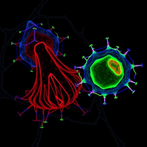
Scientists have built the first laboratory-grown fat tissue organoids that successfully recruit living T cells, recreating inside a dish the critical immune-fat dialogue that drives obesity-related metabolic disease. The new model, described in the journal iScience, allows researchers to watch regulatory T cells migrate into visceral fat spheroids within days, offering a long-sought platform to dissect why visceral fat becomes inflamed in obesity while subcutaneous fat largely resists it.
Metabolic diseases such as type 2 diabetes, fatty liver disease and cardiovascular disease represent one of the defining health challenges of the twenty-first century, and obesity sits at the center of that epidemic. Fat tissue in obesity is not metabolically inert; it is chronically inflamed, packed with neutrophils, macrophages, B cells and T cells whose interactions with adipocytes shape systemic insulin sensitivity. Among these immune players, regulatory T cells, or Tregs, have emerged as decisive guardians of fat tissue homeostasis. Visceral adipose tissue in lean mammals harbors a distinctive population of Tregs with transcriptional profiles and antigen receptor repertoires unlike those of lymphoid tissue Tregs. When these cells are experimentally depleted in living animals, adipose inflammation worsens and metabolic parameters deteriorate, with blood glucose rising and insulin sensitivity falling. Human studies mirror this picture: visceral fat Tregs are diminished in obesity and are important for maintaining glucose tolerance.
The problem has always been access. T cells resident in fat tissue can be isolated only in very low numbers from fat depots, making functional analysis of how they interact with adipocytes extraordinarily difficult. A team led by Christian Kühne and Kim Ohl at RWTH Aachen University set out to solve this by expanding an ex vivo adipose tissue organoid system the group had previously shown to be suitable for immunometabolic studies. Those earlier organoids contained mature adipocytes and some immune cells, mostly macrophages and eosinophils, but no T cells. The new work closes that gap by adding splenocytes from donor mice to fully differentiated fat organoids and letting the T cells migrate in on their own.
The protocol takes twenty days from tissue harvest to T cell-containing organoids. Stromal vascular fraction cells are first isolated from visceral fat surrounding the testes or subcutaneous fat from the inguinal region of lean male C57BL/6 mice, digested with collagenase, and briefly pre-cultured in two dimensions. The cells are then seeded at 50,000 cells per well into ultra-low attachment, U-bottom 96-well plates, where they self-assemble into spheroids within roughly three days. Adipocyte differentiation is driven by a classic cocktail of insulin, dexamethasone, rosiglitazone and IBMX, along with the thyroid hormone T3 and supporting micronutrients such as ascorbic acid, biotin and pantothenic acid. After eight days of induction and differentiation, the resulting organoids are mature adipocyte-rich spheroids ready for co-culture. One million freshly isolated splenocytes are then added to each well, and over the next two days T cells actively infiltrate the organoids.
Flow cytometry, the workhorse of the analysis, revealed a striking depot-specific pattern. When splenocytes were co-cultured with organoids made from visceral fat, CD45-positive CD3-positive T cells were readily detected inside the spheroids. Organoids derived from subcutaneous fat, by contrast, remained essentially T cell-free under unstimulated conditions. Even more intriguing, the T cells that entered visceral fat organoids contained a markedly higher proportion of FOXP3-positive Tregs, tracked with a red fluorescent reporter, than the T cells remaining in the surrounding supernatant. This suggests that visceral fat organoids preferentially recruit the regulatory subset, recapitulating the Treg-rich milieu of lean visceral fat in vivo. The infiltrating cells also showed elevated CD69 expression, although only about ten percent of them carried this early activation marker, indicating that most infiltrating T cells remain in a quiescent, tissue-resident-like state. The data further suggested that the infiltrating cells were predominantly CD4-positive, while CD8-positive cells largely stayed behind in the supernatant.
The researchers then asked what happens when the system is pushed into an inflammatory state, mimicking the conditions of obese adipose tissue. A one-hour pulse of lipopolysaccharide, or LPS, the cell wall component of E. coli that activates the innate immune receptor Toll-like receptor 4, transformed the behavior of both organoid types. Subcutaneous fat organoids, which had previously refused T cell entry, now became infiltrable, recruiting CD4-positive T cells at measurable rates. Visceral organoids showed a further, more modest increase in T cell entry. Crucially, the quality of the infiltrate differed: LPS-stimulated subcutaneous organoids contained significantly lower frequencies of Tregs than their visceral counterparts, implying that inflammation preferentially pulls in effector T cells rather than regulatory ones. This mirrors the in vivo shift, well documented in obesity research, in which CD4-positive effector T cells convert the anti-inflammatory atmosphere of lean visceral fat into a pro-inflammatory environment.
Cytokine measurements reinforced the physiological relevance of the model. Interleukin-6 secretion from visceral organoids tended to rise when splenocytes were present and climbed further after LPS stimulation, while tumor necrosis factor-alpha, a key inflammatory mediator in obese adipose tissue, increased significantly in LPS-stimulated co-cultures. The authors propose a mechanistic framework grounded in prior literature: LPS activates TLR4 on adipocytes, adipose stem cells and macrophages within the organoids, prompting the secretion of TNF-alpha and IL-6. TNF-alpha in turn induces adipose stem cells to express the chemokine CCL5, which signals through the receptor CCR5 on T cells and draws them into the tissue. Exactly how adipocyte activation orchestrates T cell migration, and why Tregs and effector T cells respond differently, are questions the team plans to pursue in this system.
The model’s design makes it unusually flexible. Because the T cells are supplied externally from splenocytes, they can be harvested from any of the vast catalogue of genetically engineered mouse strains, allowing researchers to test the effects of gene overexpression, knockdown or deficiency in the context of intact fat tissue. Drug compounds can be added directly to the culture to screen for therapeutic candidates. Standard downstream assays, including histology, flow cytometry, RNA analysis and cytokine enzyme-linked immunosorbent assays, all work with the organoids. The team also notes that human subcutaneous fat organoids have recently been generated by similar self-organization approaches, raising the prospect of a human version of this immune-fat co-culture platform built from donor material.
The authors are candid about the system’s current limits. Each organoid starts from only 50,000 stromal cells, so multiple spheroids must be pooled to harvest enough material for molecular analysis, and the excess of splenocytes in the co-culture does not perfectly reflect physiological cell ratios. The work has so far been performed exclusively with cells from male mice, even though recent studies show that visceral fat Tregs carry sex-specific gene expression programs and that two distinct Treg populations shape systemic metabolism, making replication in female mice a clear priority. LPS is also a somewhat blunt instrument for inducing inflammation, and the team intends to test more physiological stimuli such as TNF-alpha and interleukin-1 beta. Enzymatic digestion before flow cytometry can also perturb surface markers like CD4 and CD8, a technical wrinkle the group hopes to mitigate with gentler protocols and intracellular staining.
Even with these caveats, the advance addresses a genuine bottleneck. Visceral fat Tregs are rare, depot-restricted and notoriously difficult to study, yet they sit at the fulcrum of the relationship between obesity, inflammation and insulin resistance. Recent work has shown, for example, that cholesterol homeostasis is a key metabolic pathway for visceral Treg accumulation and that its disruption may explain Treg loss in obese humans and mice, a finding that could now be interrogated directly in the organoid system. A reproducible dish-based model in which fat tissue actively recruits its characteristic immune residents opens the door to mechanistic studies of T cell differentiation and migration into fat, dissection of Treg function in adipose inflammation and insulin resistance, and drug screening for new therapies targeting the immune-fat axis. In an era when metabolic disease strains health systems worldwide, a twenty-day recipe for growing immunologically complete fat tissue in a 96-well plate may prove to be a quietly powerful tool.
Cite Scienmag News
Denise Maddox. (September 7, 2026). Scientists create fat tissue organoids containing functional T cells. Scienmag. https://scienmag.com/scientists-create-fat-tissue-organoids-containing-functional-t-cells/
Denise Maddox. "Scientists create fat tissue organoids containing functional T cells." Scienmag, 7 September 2026, https://scienmag.com/scientists-create-fat-tissue-organoids-containing-functional-t-cells/. Accessed 7 September 2026.
Denise Maddox. "Scientists create fat tissue organoids containing functional T cells." Scienmag. September 7, 2026. https://scienmag.com/scientists-create-fat-tissue-organoids-containing-functional-t-cells/


