


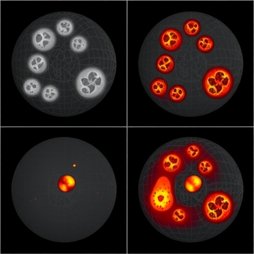
The battle against melanoma, the deadliest form of skin cancer, has just received a significant boost from an international team of researchers who have unveiled a new benchmark dataset designed to transform how artificial intelligence systems learn to detect and stage the disease. The dataset, named Mel-DEPTHS, addresses one of the most stubborn bottlenecks in computational pathology: the sheer difficulty of producing pixel-level annotated images of melanoma tissue that are accurate, consistent, and clinically meaningful. The work, published in Medical & Biological Engineering & Computing, is the product of a collaboration between computer engineers at Yildiz Technical University, dermatopathologists at Istanbul University-Cerrahpasa, and a pathologist at Memorial Sloan Kettering Cancer Center in New York, and it arrives at a moment when the demand for reliable digital pathology tools has never been greater.
Melanoma staging depends fundamentally on the precise delineation of two structures in a biopsy slide: the epidermis, the outermost layer of the skin, and the tumor itself. The depth to which tumor cells invade below the epidermis, along with features such as ulceration, determines the pathological tumor stage, or pT stage, which in turn drives decisions about surgery, sentinel lymph node biopsy, and adjuvant therapy. Yet the process of marking these boundaries pixel by pixel on whole-slide images, gigapixel-scale digital scans of entire tissue sections, is extraordinarily labor-intensive. A single slide can contain tens of billions of pixels, and even experienced dermatopathologists can disagree on exactly where the epidermis ends and the dermis begins, or where the invasive front of a tumor lies. Studies cited by the research team show that discordance in the histopathologic diagnosis of melanoma is far from rare, and that second expert reviews can change therapeutic approaches in a substantial fraction of cases. Against this backdrop, the absence of standardized, publicly available benchmarks with expert-validated ground truth has held back progress in automated melanoma staging.
Mel-DEPTHS tackles this problem head-on. The dataset comprises 50 anonymized melanoma whole-slide images digitized at 40x magnification, corresponding to a spatial resolution of 0.25 micrometers per pixel, a fidelity high enough to resolve individual nuclei and the fine architectural features that pathologists rely upon. Each image is accompanied by pixel-level masks labeling both epidermal and tumor regions, verified by dermatopathologists. Crucially, the dataset is not merely a collection of images and masks. It also carries clinical variables for each case, including invasion depth, ulceration status, and pT stage, allowing researchers to connect segmentation performance directly to the staging information that matters in the clinic. To guard against irreproducible results, the team provides fixed train and test partitions, ensuring that different laboratories evaluating their algorithms on Mel-DEPTHS are working under identical conditions and that reported results can be compared on a level playing field.
Perhaps the most innovative element of the study is the annotation strategy the researchers developed to lighten the burden on human experts. They call it Expert-Supervised Iterative Self-Training, or ESIST. The protocol begins with a pretrained segmentation model that generates initial pseudo-labels across the slides, essentially machine-generated guesses at the boundaries of epidermis and tumor. Dermatopathologists then review and refine these pseudo-labels, correcting errors and sharpening ambiguous boundaries, and the corrected labels are fed back to retrain the model. The improved model generates better pseudo-labels in the next iteration, which the experts refine again, and the cycle continues. This human-in-the-loop approach inverts the traditional workflow: instead of starting from a blank canvas and hand-drawing every pixel, experts spend their time correcting and validating machine proposals, a task that is both faster and, the team argues, less fatiguing. Self-training and pseudo-labeling techniques have become increasingly popular in semi-supervised medical image segmentation, but Mel-DEPTHS is among the first efforts to embed such a protocol into the construction of a fully expert-validated public benchmark for melanoma.
To establish how well current state-of-the-art architectures perform on this new benchmark, the researchers evaluated six leading segmentation models: UNet, UNet++, UNet3+, UPerNet, TransUNet, and ConvUNeXt. These architectures represent the major lineages of modern medical image segmentation. The UNet family, first introduced in 2015, uses an encoder-decoder design with skip connections that fuse high-resolution spatial detail from the encoder with the semantic context of the decoder; UNet++ and UNet3+ refine this idea with nested and full-scale skip connections, respectively. UPerNet, drawn from the scene-parsing literature, applies a pyramid pooling module to capture context at multiple scales. TransUNet replaces part of the convolutional encoder with a vision transformer, whose self-attention mechanism can model long-range dependencies across an image, an advantage when tumor architecture spans broad regions of tissue. ConvUNeXt adapts ideas from modern hierarchical convolutional networks for medical segmentation efficiency.
The results of the benchmark were instructive. TransUNet achieved the best overall performance across the whole-slide-level metrics, which included precision, recall, intersection over union, and Dice score, followed closely by ConvUNeXt and UPerNet. The superiority of the transformer-based model suggests that the ability to attend to global context across a slide is genuinely valuable for the fine-grained task of tracing epidermal and tumor boundaries in melanoma histopathology, where local texture cues alone may be ambiguous. The fact that the rankings were consistent across evaluation metrics strengthens confidence that these findings are not artifacts of any single measure. The team also performed three-fold cross-validation, confirming that model rankings remained stable and that the label quality produced through the ESIST protocol was robust, an important validation given that the annotations were built iteratively rather than drawn from scratch.
The significance of a well-constructed public dataset of this kind is difficult to overstate. In the broader field of artificial intelligence, benchmarks such as ImageNet catalyzed revolutions by giving researchers a common target to measure against, and medicine has seen similar dynamics with datasets for chest radiographs and organ segmentation. But histopathology of melanoma has lacked an equivalent resource at whole-slide scale with pixel-level annotations of the structures most relevant to staging. Without such a benchmark, individual groups have trained models on private collections with variable annotation standards, making it nearly impossible to tell whether published improvements reflect genuine algorithmic advances or simply differences in labeling practice. By combining expert-validated masks, clinically meaningful variables, and fixed evaluation partitions, Mel-DEPTHS creates a shared yardstick, and the authors argue it establishes both the fidelity and the diversity necessary for clinically meaningful segmentation research.
The clinical stakes are considerable. Cutaneous melanoma accounts for the majority of skin cancer deaths despite representing only a small fraction of skin cancer cases, and global burden analyses project rising incidence in the coming decades. Deep learning has already demonstrated pathologist-level performance in classifying melanoma histopathology images, and clinical-grade computational pathology systems built on weakly supervised deep learning are entering practice in other domains. Accurate automated segmentation of epidermis and tumor is a foundational step toward automating staging measurements such as Breslow thickness, the single most important prognostic factor in primary cutaneous melanoma. A system that reliably traces the epidermal surface and the deepest invaded tumor cell could, in principle, assist pathologists in measuring invasion depth consistently, flagging cases where staging is borderline, and reducing the interobserver variability that can alter treatment decisions.
The dataset is being released under a Creative Commons Attribution-NonCommercial-NoDerivatives 4.0 license, and the histopathology images are publicly accessible through the YTU Vision Research Group, making Mel-DEPTHS available to any laboratory in the world with an internet connection. The research was funded by TUSEB, the Health Institutes of Türkiye, under a Group B R&D project, and was conducted as part of the doctoral dissertation of the corresponding author, Yasemin Topuz, with ethical approval from the Non-Invasive Clinical Research Ethics Committee of Istanbul University-Cerrahpasa. The multidisciplinary authorship, spanning computer engineering, pathology, and biomedical engineering, reflects the convergence of expertise that modern computational pathology demands.
For the field of digital pathology, Mel-DEPTHS represents more than a new data resource; it embodies a workflow philosophy in which machine-generated proposals and human expertise reinforce each other rather than compete. As transformer architectures and hybrid convolutional models continue to evolve, having a standardized, clinically annotated benchmark on which to test them should accelerate the translation of laboratory algorithms into tools that genuinely support pathologists at the microscope. If the trajectory of AI-assisted diagnostics elsewhere in medicine is any guide, the iterative, expert-supervised approach demonstrated here may become a template for building high-quality annotated datasets in other cancers where pixel-level ground truth is scarce and staging precision saves lives.
Cite Scienmag News
Nathaniel Bowman. (September 7, 2026). Mel-DEPTHS: new benchmark dataset for melanoma skin and tumor segmentation. Scienmag. https://scienmag.com/mel-depths-new-benchmark-dataset-for-melanoma-skin-and-tumor-segmentation/
Nathaniel Bowman. "Mel-DEPTHS: new benchmark dataset for melanoma skin and tumor segmentation." Scienmag, 7 September 2026, https://scienmag.com/mel-depths-new-benchmark-dataset-for-melanoma-skin-and-tumor-segmentation/. Accessed 7 September 2026.
Nathaniel Bowman. "Mel-DEPTHS: new benchmark dataset for melanoma skin and tumor segmentation." Scienmag. September 7, 2026. https://scienmag.com/mel-depths-new-benchmark-dataset-for-melanoma-skin-and-tumor-segmentation/


