


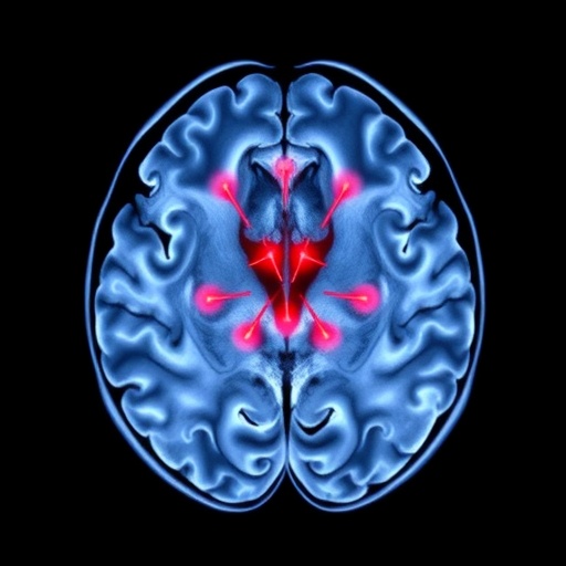
In a finding that could reshape how scientists understand and ultimately treat fear-related mental illness, researchers have used one of the world’s most powerful human brain scanners to map, with unprecedented precision, the wiring of the brain’s tiny alarm center, the locus coeruleus, in people living with anxiety and posttraumatic stress disorder. The study, published in Translational Psychiatry, reveals that this minuscule blue-colored nucleus in the brainstem communicates abnormally with two specific cortical hubs, the cingulate cortex and the insula, in ways that appear strikingly similar across two clinically distinct diagnoses. The result suggests that anxiety disorders and PTSD may share a common neural fingerprint rooted in the brain’s principal norepinephrine-producing system.
The locus coeruleus has long fascinated neuroscientists despite its diminutive size. This bilaterally paired structure contains roughly 50,000 neurons on each side of the brainstem, yet it supplies nearly all of the norepinephrine, also known as noradrenaline, to the entire cerebral cortex. Norepinephrine is central to arousal, vigilance, attention and the physiological response to threat. When a person perceives danger, the locus coeruleus fires, flooding the brain with a chemical signal that sharpens attention, quickens the heart and etches memories with emotional intensity. For decades, animal studies and pharmacological research have implicated hyperactivity of this system in the exaggerated startle, hypervigilance and intrusive memories that define PTSD and anxiety disorders. What has been missing is a direct, detailed picture of how this tiny nucleus connects to the rest of the human brain in living patients, a gap caused chiefly by the structure’s small size, its deep location and its proximity to the flow-sensitive ventricles and large vessels of the brainstem.
That gap is precisely what the new study set out to close using ultra-high field magnetic resonance imaging at 7 Tesla, roughly double the field strength of standard clinical MRI scanners. At 7 Tesla, the signal-to-noise ratio increases dramatically and the spatial resolution improves enough to visualize and delineate structures only a few millimeters across. Functional connectivity MRI, which tracks spontaneous, synchronized fluctuations in blood-oxygen-level-dependent, or BOLD, signal between brain regions while a subject rests quietly in the scanner, allowed the investigators to assess how strongly the locus coeruleus communicates with the rest of the brain without requiring any task. By carefully segmenting the locus coeruleus on individual participants’ ultra-high-resolution images, the team could measure its intrinsic functional architecture with a fidelity previously unattainable in clinical populations.
The researchers studied individuals diagnosed with anxiety disorders, individuals diagnosed with PTSD and healthy comparison participants, comparing the functional connectivity profiles of the locus coeruleus across the three groups. The central question was straightforward but technically demanding: does the brain’s norepinephrine hub engage differently with cortical and subcortical networks in people with pathological fear responses, and if so, do the patterns differ between anxiety and PTSD or do they converge?
The answer, according to the data, is convergence. In both patient groups, the locus coeruleus showed altered functional connectivity with a remarkably consistent set of regions: the cingulate cortex and the insula. The cingulate cortex, arching above the corpus callosum, is a core node of the brain’s salience and cognitive control networks, involved in detecting conflict, evaluating threat and regulating emotional responses. The insula, buried deep within the lateral sulcus, is the primary cortical representation of interoception, the sense of the body’s internal state, including heartbeat, breathlessness and visceral discomfort. Abnormal activity in both structures has repeatedly been reported in fear-based disorders, but the new findings suggest that their abnormal engagement may be driven, at least in part, by altered communication with the norepinephrine system itself.
This pattern carries significant theoretical weight. The salience network, anchored by the anterior cingulate and anterior insula, is thought to determine which internal and external stimuli capture the brain’s attention and trigger defensive responses. If the locus coeruleus, the brain’s arousal accelerator, is functionally over-wired or dysregulated in its dialogue with these regions, the result would be precisely the clinical picture seen in anxiety and PTSD: a system that assigns excessive threat salience to benign stimuli, sustains vigilance long after danger has passed and keeps the body in a state of chronic mobilization. The finding that this circuit signature appears in both disorders, despite their different diagnostic criteria and typical triggers, hints at a transdiagnostic mechanism, a shared biological substrate that cuts across the boundaries of conventional psychiatric categories.
The technical achievement underlying these results should not be understated. The locus coeruleus is notoriously difficult to image reliably. It is small enough that even slight head motion or partial-volume effects, where a single voxel, the three-dimensional picture element of an MRI scan, mixes signal from the nucleus with surrounding tissue or cerebrospinal fluid, can distort measurements. The pulsation of cerebrospinal fluid in the fourth ventricle and the nearby basilar artery introduces physiological noise exactly where researchers need a clean signal. Ultra-high field imaging mitigates many of these problems: smaller voxels reduce partial-volume contamination, and stronger gradients allow faster acquisitions that can sample the BOLD signal more thoroughly. Studies of this kind have only become feasible as 7 Tesla scanners have spread from a handful of research centers into broader use, and the new work represents one of the most direct applications of the technology to psychiatric pathophysiology to date.
Beyond its immediate anatomical findings, the study opens a window onto treatment development. Norepinephrine signaling has long been a pharmacological target in trauma-related disorders. Prazosin, an alpha-1 adrenergic antagonist, has been tested for PTSD-related nightmares with mixed results, while medications that modulate norepinephrine reuptake are mainstays in anxiety treatment. The new connectivity maps provide a potential neuroimaging biomarker: a measurable, reproducible signature of locus coeruleus, cingulate and insular communication that could be tracked before and after treatment to determine whether a given intervention actually normalizes the arousal circuitry. In a field where psychiatric diagnosis still relies heavily on self-report and clinical observation, objective neural markers of this kind are sorely needed, and functional connectivity measured at ultra-high field may offer one.
The convergent findings across anxiety and PTSD also align with a broader movement in psychiatry away from categorical diagnosis and toward dimensional, circuit-based frameworks such as the National Institute of Mental Health’s Research Domain Criteria. Rather than treating anxiety disorders and PTSD as wholly separate illnesses, the data suggest they may be viewed as points on a continuum of threat-processing dysregulation, with the locus coeruleus-norepinephrine system acting as a common engine. This does not mean the disorders are identical, and the study’s cross-sectional design cannot establish whether the connectivity alterations are a cause of symptoms, a consequence of chronic illness, or both. Longitudinal studies following at-risk individuals before the onset of disease, as well as interventions that directly modulate locus coeruleus activity, would be needed to untangle causality.
There are, as with any neuroimaging study, additional caveats worth noting. Functional connectivity reflects statistical dependence between signals, not direct anatomical wiring, and alterations in BOLD-based synchrony can arise from changes in vascular reactivity, neurotransmitter levels or neuronal coupling. Medication status, comorbid depression, prior trauma exposure and age all influence both brain connectivity and scanner data, and large, carefully phenotyped samples will be needed to confirm and extend the findings. Nevertheless, the convergence of the current results with an extensive animal literature on norepinephrine and fear, and with human task-based imaging studies of salience network dysfunction, gives the findings considerable plausibility.
What the study ultimately delivers is a sharper picture of where, in the living human brain, the machinery of pathological fear may reside, and a demonstration that 7 Tesla imaging can resolve structures once considered beyond the reach of human neuroscience. As ultra-high field scanners proliferate and analytic methods mature, the locus coeruleus may finally give up its secrets, transforming a tiny blue dot in the brainstem from an object of inference into a target of precise, circuit-level medicine for the millions of people worldwide whose lives are shadowed by anxiety and the unfinished aftermath of trauma.
Cite Scienmag News
Glenn Wilkins. (September 6, 2026). Ultra-high field MRI maps locus coeruleus connectivity in anxiety and PTSD. Scienmag. https://scienmag.com/ultra-high-field-mri-maps-locus-coeruleus-connectivity-in-anxiety-and-ptsd/
Glenn Wilkins. "Ultra-high field MRI maps locus coeruleus connectivity in anxiety and PTSD." Scienmag, 6 September 2026, https://scienmag.com/ultra-high-field-mri-maps-locus-coeruleus-connectivity-in-anxiety-and-ptsd/. Accessed 6 September 2026.
Glenn Wilkins. "Ultra-high field MRI maps locus coeruleus connectivity in anxiety and PTSD." Scienmag. September 6, 2026. https://scienmag.com/ultra-high-field-mri-maps-locus-coeruleus-connectivity-in-anxiety-and-ptsd/


