


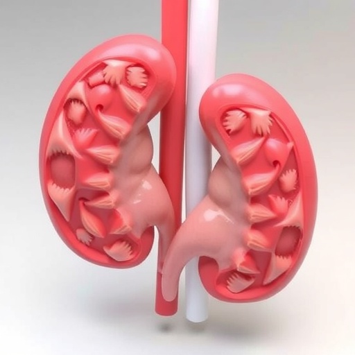
Medical students staring at flat CT scans may soon have a much better way to learn what kidney tumors actually look like in three dimensions. A new pilot study from French researchers shows that 3D-printed and virtual kidney models significantly improve medical students’ ability to assess renal tumor complexity, boosting both accuracy and speed compared with reading computed tomography images alone. The findings, published in the journal 3D Printing in Medicine, suggest that hands-on and screen-based 3D anatomical models could play a far larger role in medical education than previously assumed, and that cheaper virtual versions may be just as effective as expensive printed replicas.
The study, led by urologists and researchers at Bordeaux University Hospital in France, set out to answer a deceptively simple question: can 3D models help students move beyond memorizing anatomy and toward genuine spatial understanding of kidney tumors? Radiological training has long been underrepresented in medical curricula, even though interpreting cross-sectional imaging is a skill nearly every practicing clinician needs. Meanwhile, kidney cancer management depends heavily on accurate imaging and on standardized tumor complexity scores, such as the R.E.N.A.L. nephrometry score and the PADUA classification, which help surgeons decide whether a patient’s tumor can be safely removed while preserving the kidney. The researchers reasoned that if students could improve at completing these scores by using 3D models, the models would be doing more than making anatomy prettier; they would be sharpening skills with direct clinical relevance.
To build the learning tools, the team selected three kidney tumor cases from patients who had undergone robotic partial nephrectomy, deliberately choosing examples of low, intermediate, and high anatomical complexity. The patients’ CT scans were anonymized and segmented using Synapse 3D software from FUJIFILM, a platform capable of delineating individual anatomical structures from imaging data. A urologist performed the initial segmentation, generating digital models that included the tumor, the healthy renal cortex, the arterial and venous vasculature, and the urinary collecting system. These digital reconstructions were then cross-verified against the original CT scans and refined for anatomical accuracy by a senior urologist and a senior radiologist, an essential quality control step, since even small errors in a printed teaching model could quietly teach students the wrong anatomy.
The segmented models were exported as standard STL files and processed for additive manufacturing. For the physical versions, the team used a Stratasys J750 3D printer, a machine that relies on photopolymer material jetting technology to produce high-resolution, multi-material objects. This approach allowed the researchers to print different structures in different materials and colors within a single model, and to use translucent resin for the renal cortex so that the hilar structures, where the vessels and ureter enter and exit the kidney, remained visible through the outer shell. The printed models were revalidated against the original CT scans by two urologists and a senior radiologist before entering the classroom.
Twenty-three fifth-year medical students were then recruited and randomized into three groups. Seven students received only the anonymized CT scans and a standard viewer, forming the CT-only group. Nine students worked with the CT scans plus an interactive 3D virtual model that they could rotate and manipulate on the same computer, forming the 3DV group. Seven students had access to the physical 3D-printed models during interpretation, forming the 3DP group. All participants had previous exposure to CT interpretation through earlier clinical rotations, and baseline questionnaires confirmed that the groups did not differ significantly in medical, radiological, or urological experience, meaning the results could reasonably be attributed to the learning tools rather than to unequal starting knowledge.
Each student independently interpreted the same three clinical cases, completing questionnaires for every case that incorporated items drawn directly from the PADUA and RENAL complexity scoring systems. Two urologists and a radiologist also added six supplementary questions per case, targeting anatomical details with clinical implications. Every correct answer earned one point, for a potential total of 17 points per case. The students were timed throughout, and the entire exercise concluded with a feedback session, followed by an anonymous satisfaction survey. Because the sample sizes were small and unequal, the researchers used non-parametric Mann-Whitney tests for all comparisons rather than assuming normally distributed data.
The results were striking. Accuracy in completing the complexity scores reached a median of 91 percent in the virtual model group and 91 percent in the printed model group, compared with only 73 percent in the CT-only group, a difference that was statistically significant for both 3D groups. Students using 3D tools also worked faster. Mean completion time was about 11 minutes in the CT-only group, 9.4 minutes with the virtual model, and just 7.1 minutes with the printed model, with both 3D groups significantly outperforming the CT-only group on time as well. Total scores combining complexity assessments and anatomical questions told the same story: roughly 62 percent for the CT-only group, versus 81 percent for the virtual group and 83 percent for the printed group. Crucially, there was no statistically significant difference between the virtual and printed model groups on any measure.
Student enthusiasm matched the quantitative gains. Median satisfaction with the 3D-printed model as a radiology teaching tool was 80 percent, the perceived benefit for understanding anatomy hit a full 100 percent, and 90 percent of respondents said similar tools should be developed for other organs and medical disciplines. The models also earned high marks for clarifying the relationship between the tumor and the kidney, an understanding that matters enormously when surgeons are deciding whether a tumor can be excised without sacrificing renal function.
The authors are careful to frame what the improvement actually represents. The 3D groups did not acquire formal radiological expertise; rather, the models enhanced spatial understanding and task performance, helping students search for and grasp anatomical relationships within the scans. That distinction matters, but the underlying skill, reading cross-sectional imaging accurately, is universally valuable. Most medical students will not become urologists, yet nearly all will need to interpret CT images at some point in their careers, whatever specialty they choose. The researchers argue that the 3D approach bridges the gap between static textbook anatomy and the dynamic, three-dimensional reasoning that real radiological interpretation demands.
The equivalence of virtual and physical models carries perhaps the biggest practical implication. Earlier work, including a study by Wake and colleagues on patient education, had suggested printed models might outperform virtual ones. The Bordeaux team suspects the difference is generational: today’s medical students are fluent with digital 3D visualization tools, virtual reality, and augmented reality, so a screen-based interactive model may convey as much spatial information to them as a physical object. That finding has immediate budgetary consequences, since printed kidney models typically cost between 150 and 500 dollars each, while virtual models are essentially free to reproduce and distribute once the segmentation work is done. For medical schools weighing how to modernize their curricula, the study suggests virtual models could deliver comparable educational benefits at a fraction of the cost.
The study builds on a growing literature showing 3D-printed models succeeding in other contexts: improving trainee understanding of kidney tumor anatomy, helping patients grasp their own surgical plans, and even outperforming cadaveric materials in randomized trials of cardiac anatomy education. What distinguishes the new work is its focus on undergraduate learners rather than surgical residents, and its use of validated complexity scoring as an objective outcome measure rather than relying on opinion surveys. The researchers also emphasize rigorous accuracy verification of their models, following the same methodological standards they described in earlier work on 3D-printed model accuracy for robotic partial nephrectomy planning.
Limitations remain, and the authors are candid about them. This was a small pilot involving only three tumor cases and 23 students, with unequal group sizes that limit statistical power. The design did not measure pre- and post-intervention changes in CT interpretation ability, so long-term learning effects are unknown, and restricting cases to a single organ system leaves open the question of how well the approach generalizes. The team calls for larger randomized controlled trials across multiple disciplines and organ systems to confirm and extend the findings.
Even with those caveats, the implications are hard to ignore. Kidney cancer is a major global health concern, and nephron-sparing surgery depends on precisely the kind of spatial reasoning these models cultivate. Tools that demonstrably improve students’ accuracy, speed, and confidence in interpreting renal CT scans, at low cost and with high learner satisfaction, address a genuine gap in medical training. If the results hold up at scale, the anatomy lab of the future may include far fewer flat scans and far more objects that students can hold in their hands, or at least spin on a screen, before they ever meet a patient.
Cite Scienmag News
Nathaniel Bowman. (September 6, 2026). 3D printed kidney models help students understand renal tumor anatomy. Scienmag. https://scienmag.com/3d-printed-kidney-models-help-students-understand-renal-tumor-anatomy/
Nathaniel Bowman. "3D printed kidney models help students understand renal tumor anatomy." Scienmag, 6 September 2026, https://scienmag.com/3d-printed-kidney-models-help-students-understand-renal-tumor-anatomy/. Accessed 6 September 2026.
Nathaniel Bowman. "3D printed kidney models help students understand renal tumor anatomy." Scienmag. September 6, 2026. https://scienmag.com/3d-printed-kidney-models-help-students-understand-renal-tumor-anatomy/


