


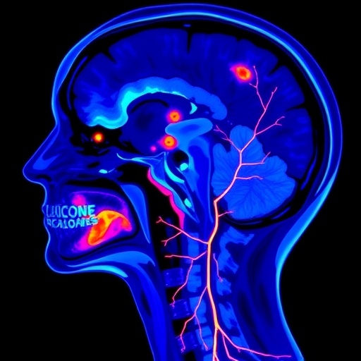
Cancer therapy saves lives, but it can quietly injure the blood vessels that keep the rest of the body alive. A new study from the University of California Davis Health offers the clearest whole-body picture yet of that hidden toll, showing that patients treated for head and neck cancer develop measurable inflammation in arteries far beyond the irradiated region, and that this inflammation scales with the dose of radiation they received. Using a state-of-the-art total-body PET scanner and an unconventional delayed imaging protocol, the research team captured the vascular signature of concurrent chemoradiotherapy in unprecedented detail, raising the prospect of a new imaging biomarker for cardiovascular risk in cancer survivors.
The findings, published in the European Journal of Nuclear Medicine and Molecular Imaging, center on a tracer familiar to nuclear medicine physicians everywhere: 2-[18F]fluoro-2-deoxy-D-glucose, or FDG. This radioactive glucose analogue accumulates in metabolically active cells, which is why it has long been the workhorse of oncological PET imaging. But metabolically active cells include not only tumors but also inflammatory cells such as macrophages embedded within arterial walls. When vascular inflammation flares, FDG uptake in arteries rises, and that signal can be quantified. Clinicians have exploited this property for more than a decade to study atherosclerosis, but conventional PET scanners, with their narrow axial fields of view, can only image short segments of the vasculature at a time, and their limited sensitivity makes prolonged imaging protocols impractical.
Enter the uEXPLORER, the world’s first total-body PET/CT scanner, installed at UC Davis. With an axial field of view that spans the entire body in a single bed position and a sensitivity roughly an order of magnitude higher than conventional scanners, total-body PET changes the mathematics of molecular imaging. It allows researchers to image the entire arterial tree from the carotid arteries at the top of the neck down to the infrarenal abdominal aorta in one acquisition, and to extend the uptake time well beyond the standard hour without increasing the injected radiation dose to prohibitive levels. In this study, the team exploited exactly that capability: patients received FDG injection and then waited a full two hours before scanning, rather than the customary sixty minutes.
The rationale for the delay is rooted in tracer kinetics. FDG circulating in the blood pool creates a bright background that can obscure or confound measurements of uptake in the thin walls of arteries, which sit perilously close to flowing blood. As time passes, blood-pool activity clears while activity trapped in inflammatory cells remains, improving the contrast between vessel wall and blood. Prior work has shown that longer circulation times improve the reproducibility and diagnostic quality of vascular FDG measurements, and the European Association of Nuclear Medicine has acknowledged the importance of uptake timing in its position papers on atherosclerosis imaging. By combining delayed imaging with total-body sensitivity, the UC Davis team aimed to maximize the signal-to-noise ratio of vascular inflammation measurements across the whole arterial system.
Twenty-four patients with head and neck cancer underwent 2-hour delayed total-body PET/CT both before and three months after concurrent chemoradiotherapy, the standard of care for locally advanced disease in this patient population. The researchers evaluated vascular inflammation across eleven arterial segments, including the carotid arteries, subclavian arteries, and the abdominal aorta. For each image slice, they calculated the mean standardized uptake value, or SUVmean, a measure of tracer concentration corrected for injected dose and body weight. To account for individual variation in blood-pool activity, they normalized each arterial SUVmean to the SUVmean of the superior vena cava, a large central vein that serves as an internal blood-pool reference, thereby deriving a target-to-background ratio known as TBRmean. Segmental averages of these values, termed avgTBRmean, were then compared before and after treatment.
The results were striking. Following chemoradiotherapy, avgTBRmean increased in every one of the eleven arterial segments examined, and the increases were statistically significant in several key vessels. The left carotid artery showed a rise in TBR from 1.20 to 1.32, and the right carotid from 1.24 to 1.34, both highly significant findings given the carotids’ role in supplying blood to the brain. The subclavian arteries also showed significant increases, from 1.14 to 1.20 on the left and 1.10 to 1.18 on the right, while the infrarenal abdominal aorta rose from 0.96 to 1.08. That a vessel as distant from the head and neck as the abdominal aorta should show a significant uptick in inflammatory signal suggests that the biological effects of treatment are not confined to the radiation field.
Crucially, the magnitude of vascular inflammation correlated with the radiation dose delivered. The mean radiotherapy dose received by each arterial segment correlated both with post-treatment avgTBRmean, with a correlation coefficient of 0.289, and with the relative change in TBR from baseline, with a coefficient of 0.251. These correlations, while statistically significant across the pooled segment data, paint a dose-response picture that is difficult to explain by chance: the more radiation an artery received, the more its inflammatory signal climbed. A reassuring technical detail also emerged from the analysis. The SUVmean of the superior vena cava, the internal reference, was essentially unchanged between the pre- and post-treatment scans, indicating that the arterial increases were not an artifact of altered systemic tracer distribution or changing blood-pool activity.
The clinical stakes of these measurements are considerable. Epidemiological studies have long documented that survivors of head and neck cancer face elevated risks of myocardial infarction, stroke, and carotid stenosis in the years and decades after radiotherapy, and large analyses of breast cancer radiotherapy have established that even modest doses to the heart translate into incremental ischemic risk. Data from atomic bomb survivors further demonstrate that radiation exposure accelerates atherosclerosis in a dose-dependent manner. Chemotherapy agents, including the platinum compounds frequently combined with radiation in head and neck protocols, carry their own vascular toxicities, potentiating the inflammatory milieu. What has been missing is a practical, quantitative method to observe this process as it unfolds in living patients, rather than waiting for a stenosis to appear on an ultrasound or a stroke to occur.
That is where the new biomarker shows early promise. Among the patients in the cohort, those who went on to experience cardiovascular-related events during follow-up had, on average, a substantially larger relative increase in vascular FDG uptake after treatment than those who did not: 12.1 percent versus 4.5 percent, a statistically significant difference. In other words, the patients whose arteries responded most vigorously to treatment, as measured by delayed total-body PET, were the ones most likely to develop cardiovascular complications. This finding hints that the post-treatment change in vascular FDG uptake could serve as an early warning signal, identifying survivors who might benefit from intensified cardiovascular surveillance, statin therapy, or other preventive measures long before structural disease becomes apparent on conventional imaging.
The investigators are careful, however, not to overstate the case. When the analysis was adjusted for age, the association between the relative TBR change and cardiovascular events lost its statistical independence, an unsurprising outcome in a cohort of only twenty-four patients, given that both vascular inflammation and cardiovascular risk rise steeply with age. Larger studies with longer follow-up will be needed to determine whether the imaging biomarker retains prognostic value once traditional risk factors are accounted for. The study is also exploratory in its event analysis by design, and the authors explicitly frame their conclusion as warranting validation rather than establishing clinical practice. Still, the consistency of the dose-response relationship and the uniformity of the arterial response across all eleven segments lend biological credibility to the signal.
Beyond its immediate clinical implications, the study showcases what delayed total-body FDG PET makes possible in molecular cardiovascular imaging. The ability to survey the entire arterial tree simultaneously, with the improved contrast afforded by two-hour uptake times and the internal normalization to a stable venous reference, provides a template for future investigations of therapy-induced vascular injury, whether from radiation, chemotherapy, or emerging immunotherapies that have themselves been linked to arterial inflammation. For the growing population of cancer survivors, now numbering in the tens of millions worldwide, the prospect of quantifying and monitoring the cardiovascular consequences of curative treatment offers a path toward interventions that could preserve not just cancer-free years but healthy ones. The work was supported by the National Cancer Institute through the In Vivo Translational Imaging Shared Resources, the Fred and Julia Rusch Foundation for Nuclear Medicine Research and Education, and a fellowship gift from United Imaging Health, and the study was approved by the institutional review board of UC Davis Health with written informed consent obtained from all participants.
Cite Scienmag News
Nathaniel Bowman. (September 6, 2026). Delayed FDG PET/CT reveals systemic vascular inflammation after head and neck chemoradiotherapy. Scienmag. https://scienmag.com/delayed-fdg-pet-ct-reveals-systemic-vascular-inflammation-after-head-and-neck-chemoradiotherapy/
Nathaniel Bowman. "Delayed FDG PET/CT reveals systemic vascular inflammation after head and neck chemoradiotherapy." Scienmag, 6 September 2026, https://scienmag.com/delayed-fdg-pet-ct-reveals-systemic-vascular-inflammation-after-head-and-neck-chemoradiotherapy/. Accessed 6 September 2026.
Nathaniel Bowman. "Delayed FDG PET/CT reveals systemic vascular inflammation after head and neck chemoradiotherapy." Scienmag. September 6, 2026. https://scienmag.com/delayed-fdg-pet-ct-reveals-systemic-vascular-inflammation-after-head-and-neck-chemoradiotherapy/


