


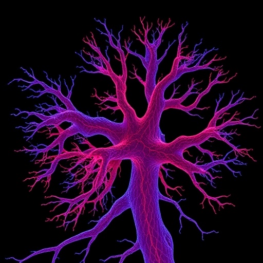
The intricate branching architecture of neurons has long fascinated neuroscientists, not only for its beauty but because it holds the key to understanding how the brain wires itself, how it malfunctions in disease, and how candidate drugs reshape neural circuits. Yet measuring that architecture rigorously has remained stubbornly difficult, particularly in the dense neuronal cultures that serve as the workhorses of modern neuropharmacology. When neurites from hundreds of neurons overlap under the microscope and background signal blurs the boundaries between cells, the traditional gold standard—painstaking manual tracing of individual arbors—becomes unreliable, slow, and often impossible. A new study published in the journal Neuroinformatics offers a compelling alternative, demonstrating that fractal mathematics can capture drug-induced changes in neuronal structure with a sensitivity that classical morphometric tools struggle to match.
The research, led by Izhak Michaelevski of Ariel University together with colleagues including Angela Asir RV, Sirish Karri, Marina Kabirova, and Michael Firer, set out to test whether two scale-invariant descriptors—fractal dimension and lacunarity—could serve as robust, global measures of neurite complexity in crowded primary hippocampal cultures. Fractal dimension, abbreviated FD, quantifies how thoroughly a structure fills space. A simple line has a fractal dimension of 1, a filled plane 2, and a densely branched neuronal arbor falls somewhere in between: the more elaborate and space-filling the branching, the closer the value creeps toward 2. Lacunarity, or LAC, complements this by measuring heterogeneity—gaps, clustering, and unevenness in how the structure is distributed across spatial scales. Together, the two parameters provide a compact statistical fingerprint of an entire neuronal network, one that does not require resolving which branch belongs to which cell.
To put the method through its paces, the team treated cultured hippocampal neurons with imatinib, the well-known Abl tyrosine kinase inhibitor best known clinically as a leukemia drug. Abl family kinases are increasingly recognized as regulators of the neuronal cytoskeleton, influencing actin dynamics, dendritic branching, and synaptic architecture, and imatinib served here as a reliable chemical tool to induce controlled cytoskeletal remodeling. The researchers imaged the treated and untreated cultures using standardized confocal microscopy, converted the images to binary representations under rigorously controlled conditions, and then skeletonized the neurite networks—reducing each arbor to its branching backbone—before computing the fractal parameters. This skeletonization step is critical: by stripping away branch thickness and focusing on topology, it allows fractal measures to reflect the true geometric complexity of the network rather than staining intensity or out-of-focus blur.
The results were striking. Imatinib treatment drove the mean fractal dimension down from approximately 1.56 in vehicle-treated control cultures to approximately 1.36 in treated cultures, a difference the authors report at a significance level of p < 0.001. That drop of roughly 0.2 in fractal dimension represents a substantial loss of multiscale complexity—essentially, the neurite networks became simpler, less elaborate, and less capable of filling two-dimensional space with fine branching. At the same time, lacunarity increased, signaling that the remaining structure was more spatially heterogeneous, with larger gaps and more irregular clustering of processes. In plain terms, the drug did not merely prune the neurons’ branches uniformly; it made the network both simpler and messier at the same time, a combined signature that a single classical measurement would struggle to convey.
Indeed, the contrast with conventional morphometry is where the study delivers its most important methodological message. When the team ran classical analyses—counting branch points, measuring primary neurite lengths, performing Sholl-style assessments—they uncovered a phenotypically mixed picture. Imatinib caused primary neurites to elongate while simultaneously reducing branching. These two effects pull in opposite directions on standard metrics, and they partially compensated for one another, obscuring the overall change in complexity when any single parameter was examined in isolation. A researcher relying on neurite length alone might conclude the drug promotes outgrowth; one relying on branch counts might conclude the opposite. Fractal dimension and lacunarity, by contrast, integrated these opposing contributions into a single coherent signal of reduced complexity and increased heterogeneity.
The authors did not stop at a pair of summary statistics. Recognizing that neuronal morphology is inherently multivariate, they constructed a composite Neurite Complexity Index, or NCI, that blends branching density, neurite length, and distribution-sensitive descriptors into one integrated score. The index performed with remarkable explanatory power, achieving a coefficient of determination of approximately 0.98 and a highly significant overall model fit (F(4,106) = 89.6, p < 1 × 10⁻¹⁸⁴). Crucially, fractal dimension converged strongly with this composite index across bootstrap resampling analyses, meaning that the simple fractal measurement captured essentially the same structural information as the far more elaborate multivariate construct. Clustering and discriminant analyses further confirmed that fractal measures possessed high discriminatory power, reliably separating drug-treated cultures from controls.
The implications extend well beyond this particular drug-neuron pairing. Dense cultures are the norm, not the exception, in high-throughput drug screening, disease modeling, and neurotoxicity testing. Manual tracing simply does not scale to thousands of wells, and automated tracing algorithms—which must untangle overlapping arbors pixel by pixel—remain error-prone precisely where cultures are most crowded. Fractal analysis sidesteps the problem entirely by operating on the network as a whole. Because fractal dimension is scale-invariant, it does not depend on magnification in the way that counts of branches or segment lengths do, making it unusually tolerant of the imaging variability that plagues multi-lab studies. The approach also aligns with a growing body of literature: fractal measures have been applied to retinal vasculature, cytoskeletal changes in osteoblasts, and, intriguingly, to neuroimaging in neurodegenerative disease, where reductions in brain fractal dimension have been reported in Alzheimer’s and Parkinson’s disease.
There is also a conceptual bridge to how neurons actually work. Earlier work from fractal-minded neuroscientists has suggested that neuronal arbors are not fractal by accident—that exploiting fractal geometry across a limited range of scales may help neurons optimize connectivity with minimal wiring cost. If neurons natively tune their arbors toward certain fractal properties, then shifts in fractal dimension and lacunarity may not merely be convenient readouts but may directly reflect biologically meaningful changes in how neurons explore space and form connections. The imatinib results fit neatly into this framework: disrupting Abl kinase signaling, which is known to regulate F-actin-dependent processes and dendritic branch maintenance, produced exactly the kind of coordinated simplification that one would expect if the kinase normally helps maintain the arbor’s fractal architecture.
The authors are appropriately measured in their claims. They frame fractal analysis as a sensitive and compact framework within the present experimental context for detecting treatment-associated structural changes in dense neuronal cultures—language that signals both enthusiasm and an awareness of scope. Fractal measures are global summaries; they cannot tell you which specific branch retracted or whether a particular subtype of neuron responded differently. Data availability is also handled pragmatically: processed image datasets, extracted morphometric parameters, and fractal analysis outputs are available from the corresponding author upon reasonable request, with all statistical analysis scripts supplied as supplementary material—an increasingly important transparency practice given that fractal results can depend on thresholding and skeletonization choices.
Still, the practical appeal is hard to overstate. A pipeline built on standardized binarized confocal images and skeletonized representations is inherently automatable, and the study’s demonstration that a single fractal dimension value tracks so closely with a sophisticated composite index suggests that screening workflows could trade layers of morphometric bookkeeping for one or two numbers per well without losing analytical power. For laboratories studying neurodegenerative and psychiatric disorders—fields where kinase inhibitors, amyloid peptides, and cytoskeletal remodeling intersect—the ability to quantify neurite complexity quickly, reproducibly, and in the dense cultures that best mimic tissue crowding could accelerate both basic discovery and preclinical testing. The study does not claim that fractal analysis should replace classical morphometry everywhere; rather, it shows that when cultures become too tangled to trace and single metrics too ambiguous to trust, the mathematics of fractals offers a way to see the forest, the trees, and the gaps between them all at once.
Cite Scienmag News
Ophelia Keating. (September 5, 2026). Fractal Analysis Tracks Structural Changes in Cultured Neurons. Scienmag. https://scienmag.com/fractal-analysis-tracks-structural-changes-in-cultured-neurons/
Ophelia Keating. "Fractal Analysis Tracks Structural Changes in Cultured Neurons." Scienmag, 5 September 2026, https://scienmag.com/fractal-analysis-tracks-structural-changes-in-cultured-neurons/. Accessed 5 September 2026.
Ophelia Keating. "Fractal Analysis Tracks Structural Changes in Cultured Neurons." Scienmag. September 5, 2026. https://scienmag.com/fractal-analysis-tracks-structural-changes-in-cultured-neurons/


