


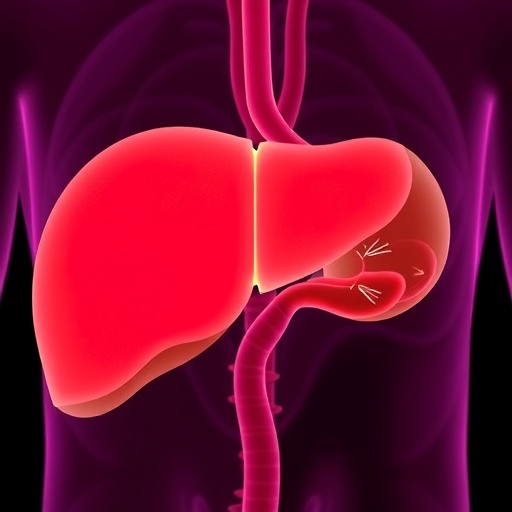
A bile acid long associated with liver damage may have a much narrower—and potentially useful—side to its biological identity. In a new study published in Life Metabolism, researchers report that carefully calibrated doses of lithocholic acid, or LCA, reduced fatty liver in mice and cynomolgus macaques without causing detectable liver toxicity. The findings suggest that the compound’s effects depend less on whether LCA is inherently harmful or beneficial than on how much reaches the bloodstream and liver.
LCA is a secondary bile acid produced when intestinal microorganisms transform primary bile acids. It participates in bile acid metabolism and can influence cellular signaling, energy balance, and inflammation. At high concentrations, however, LCA has been linked to cholestasis, impaired bile flow, obstruction of bile ducts, hepatocyte injury, and cell death. This apparent contradiction has complicated efforts to explore LCA as a possible therapy. Earlier work had shown that LCA levels rise during caloric restriction, a dietary intervention associated with longer lifespan and improved metabolic health in several organisms.
Caloric restriction is thought to activate a network of metabolic responses that improve glucose handling, mitochondrial function, stress resistance, and tissue maintenance. LCA has been proposed as one of the circulating molecules that may help transmit some of these benefits. In animal studies, the bile acid has been associated with increased muscle NAD+ levels, improved grip strength and endurance in aged mice, and longer lifespan in nematodes and fruit flies. Yet the doses required to produce such effects must be distinguished from the much higher exposures known to damage the liver.
To investigate this dose boundary, a team led by Sheng-Cai Lin of Henan University and Xiamen University first studied obese mice. The animals received LCA in their drinking water at a concentration of 1 gram per liter, producing blood levels of approximately 1 micromole per liter. That exposure was designed to resemble the concentration observed during caloric restriction rather than the substantially higher levels used in toxicology experiments. After four weeks, the mice had lower hepatic triglyceride content, reduced fatty liver, and improved glucose metabolism.
The researchers also examined the molecular pathway behind the response. Liver-specific knockout mice lacking AMPKα did not receive the same metabolic benefits, implicating AMP-activated protein kinase as a central mediator. AMPK is an energy-sensing enzyme that becomes active when cellular energy supplies are limited. Once activated, it can suppress energy-intensive processes such as lipid synthesis while promoting fatty-acid oxidation and other pathways that help restore energy balance. The results indicate that low-dose LCA may improve liver metabolism through this energy-sensing system.
The safety picture changed sharply when the dose was increased. At 250 milligrams per kilogram per day, hepatic LCA concentrations reached about 14 micromoles per liter, and the mice developed clear signs of liver injury. This contrast provided direct evidence for a dose-dependent safety window: concentrations near those associated with caloric restriction appeared beneficial, while substantially higher exposure became toxic. The distinction is particularly important because bile acids can accumulate in the liver and exert effects that are not predicted simply by the administered dose.
The team next tested LCA in cynomolgus macaques with fatty liver. Translating doses from rodents to primates proved more complicated than expected. The monkeys rejected the formulation used in the mouse experiments, so the researchers developed a phospholipid-coated preparation suspended in fish oil. When they administered a mouse-equivalent dose calculated by body-surface-area conversion—9.6 milligrams per kilogram—the animals’ serum LCA concentrations rose above 6 micromoles per liter. Within one week, alanine aminotransferase and aspartate aminotransferase, enzymes commonly used to detect liver injury, increased significantly.
That result demonstrated why standard interspecies dose conversion can be unreliable for compounds whose absorption, metabolism, and circulation differ between animals. The researchers therefore conducted a dose-titration study in the macaques and identified two lower regimens: 0.25 milligrams per kilogram and 0.5 milligrams per kilogram, administered twice daily. These schedules maintained steady-state blood concentrations of approximately 0.8 to 1 micromole per liter, close to the target range observed in the mouse experiments, without producing biochemical evidence of liver damage.
After 13 weeks, macaques receiving either low-dose regimen showed significant histological improvement in hepatic steatosis, the abnormal accumulation of fat inside liver cells. Their body weight, blood lipids, and glucose levels remained stable, while ALT, AST, creatinine, and blood counts showed no treatment-related abnormalities. The findings do not establish that LCA is ready for human use, and the study involved early-stage fatty liver rather than advanced disease with severe hyperglycemia or hypertriglyceridemia. Longer studies will be needed to assess tissue distribution, sex-related differences, chronic toxicity, and responses in more advanced models. Nevertheless, the work provides the first reported evidence in a non-human primate that a carefully controlled, caloric-restriction-like concentration of LCA may alleviate fatty liver through hepatic AMPK activation without detectable toxicity.
News Publication Date: 23-Jul-2026
Web References: https://doi.org/10.1093/lifemeta/loag023
References: Life Metabolism, DOI: 10.1093/lifemeta/loag023
Cite Scienmag News
Daisy Hatcher. (August 6, 2026). Lithocholic acid eases fatty liver disease in mice and nonhuman primates. Scienmag. https://scienmag.com/lithocholic-acid-eases-fatty-liver-disease-in-mice-and-nonhuman-primates/
Daisy Hatcher. "Lithocholic acid eases fatty liver disease in mice and nonhuman primates." Scienmag, 6 August 2026, https://scienmag.com/lithocholic-acid-eases-fatty-liver-disease-in-mice-and-nonhuman-primates/. Accessed 4 September 2026.
Daisy Hatcher. "Lithocholic acid eases fatty liver disease in mice and nonhuman primates." Scienmag. August 6, 2026. https://scienmag.com/lithocholic-acid-eases-fatty-liver-disease-in-mice-and-nonhuman-primates/


