


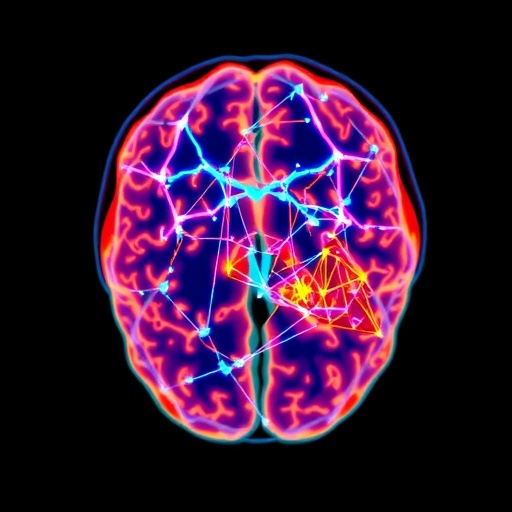
A new study led by researchers at Rutgers University and Yale University uses specialized positron emission tomography (PET) to measure synaptic connections directly in the living human brain, offering fresh clues to the biological basis of schizophrenia. Published in Molecular Psychiatry, the work moves beyond conventional imaging by targeting the density of synapses—small contact points where neurons communicate.
Synapses coordinate neural circuits that support thought, emotion, and memory. In schizophrenia, disruptions to these connections have long been suspected, yet the detailed spatial pattern of synaptic loss in living people has been difficult to observe. Standard MRI scans reveal brain size and structure, but they cannot specifically quantify synapses.
The study enrolled 122 participants, including 29 individuals diagnosed with schizophrenia. Using synaptic density PET imaging and a large dataset for this technique, the researchers compared synaptic connection levels across the brain. Results showed a pronounced and widespread reduction in synaptic density in people with schizophrenia relative to healthy participants.
The pattern of loss was not uniform. Multiple regions linked to cognition and affect—such as frontal and temporal areas, as well as brain systems involved in memory and emotion—showed significant decreases. The left hemisphere was substantially more affected than the right, indicating lateralized vulnerability rather than a purely global effect.
Importantly, the team found that synaptic loss patterns differed from MRI-detected volume alterations. This suggests schizophrenia may involve at least two partially distinct biological processes: one affecting synaptic connectivity and another influencing gross brain structure.
To understand why certain regions may be more vulnerable, the researchers examined the relationship between synaptic loss and receptor-rich molecular landscapes. Areas normally enriched in neurotransmitter receptors—specifically serotonin, gamma-aminobutyric acid (GABA), and glutamate—tended to show the greatest synaptic reductions. The findings support the idea that molecular “fitness” varies across brain regions, shaping where damage emerges.
The researchers then used computer simulations to model how synaptic loss could spread through the brain’s structural network. These analyses pointed to a likely starting region in the left frontal lobe, from which disruption may propagate to connected areas.
“These findings suggest that in schizophrenia, synaptic loss is not random,” said first author Sidhant Chopra. “Rather, it follows the brain’s molecular and connectivity architecture,” he added, implying that synaptic vulnerability may be predictable.
Senior author Avram Holmes emphasized the clinical implications: detailed mapping could help identify where interventions might preserve or restore synaptic function. The researchers propose that future longitudinal studies will clarify how synaptic loss unfolds over time and how it responds to treatments.
Overall, the work reframes schizophrenia biology as a network- and molecule-guided process, paving the way toward more precise and potentially personalized therapeutic strategies aimed at synapse protection and recovery.
Cite Scienmag News
Cassandra Pierce. (July 15, 2026). Imaging Study Finds Widespread Brain Connectivity Loss in Schizophrenia. Scienmag. https://scienmag.com/imaging-study-finds-widespread-brain-connectivity-loss-in-schizophrenia/
Cassandra Pierce. "Imaging Study Finds Widespread Brain Connectivity Loss in Schizophrenia." Scienmag, 15 July 2026, https://scienmag.com/imaging-study-finds-widespread-brain-connectivity-loss-in-schizophrenia/. Accessed 3 September 2026.
Cassandra Pierce. "Imaging Study Finds Widespread Brain Connectivity Loss in Schizophrenia." Scienmag. July 15, 2026. https://scienmag.com/imaging-study-finds-widespread-brain-connectivity-loss-in-schizophrenia/


