In a groundbreaking advancement poised to transform the study of membrane proteins, scientists from Durham University, in collaboration with Jagiellonian University in Poland, have engineered a pioneering nanoscale platform that enables the precise capture and spatial organization of these critical biological molecules. Membrane proteins, embedded within the phospholipid bilayer of cellular membranes, function as essential gatekeepers, regulating the influx and efflux of signals and various substances crucial to cellular function. Their indispensable roles in processes such as signal transduction, molecular transport, and cellular communication make them prime targets for therapeutic intervention. However, their intrinsic fragility and the complexity of their native lipid environments have historically rendered them elusive to detailed structural and functional analyses.
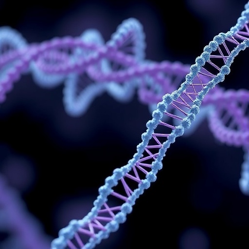
Addressing these formidable challenges, the interdisciplinary team introduced an innovative approach that synergizes the principles of DNA nanotechnology with membrane-mimetic systems. Central to their method is the construction of nanoscale DNA rings synthesized via DNA origami—a technique that exploits the predictable base-pairing of DNA strands to fold and assemble bespoke three-dimensional structures with nanometer precision. These DNA nano-rings act as scaffolding frameworks capable of spatially confining nanodiscs, which themselves are sacrificially engineered lipid bilayer patches stabilized by membrane scaffold proteins or synthetic polymers, capable of harboring individual membrane protein molecules in a native-like lipid environment.
The resulting hybrid system, named DNA-Origami-Constrained Nanodiscs (DOC-NDs), represents a quantum leap in the controlled study of membrane proteins. Unlike conventional methods that often compromise protein orientation or integrity, DOC-NDs maintain the protein’s functional conformation while rigidly positioning it within the DNA constraining ring. This architecture not only preserves the protein’s accessibility for biochemical interrogation but also significantly enhances experimental reproducibility by standardizing molecular orientation and spacing. Recent experiments demonstrated that the majority of DNA origami rings efficiently sequester nanodiscs, frequently encapsulating only a single membrane protein per construct, thereby facilitating high-resolution studies where molecular heterogeneity has historically been a significant confounding factor.
One of the most notable technical breakthroughs lies in the system’s capacity to dictate membrane protein orientation within the DNA ring. Protein orientation is crucial for accurately deciphering functional mechanisms, especially in signal transduction where the extracellular and intracellular domains must be distinctly situated. Through meticulous design, the researchers imparted directional cues that influence the insertion and presentation of proteins relative to the DNA scaffold. This level of spatial control paves the way for superior imaging techniques, most notably cryo-electron microscopy (cryo-EM), where consistent molecular orientation dramatically improves the resolution and interpretability of structural data.
Professor Jonathan Heddle, a lead investigator of the project, articulated the conceptual innovation behind this technology as a sophisticated confluence of biological molecules: “We have intricately combined protein, DNA, and lipid components to fabricate a nanoscale system that operates with unprecedented precision.” This interdisciplinary amalgamation underscores the transformative potential of merging synthetic biology and nanotechnology to tackle longstanding obstacles in membrane protein research.
From a functional perspective, DOC-NDs hold promise not only for static structural studies but also for dynamic investigations into membrane protein behaviors in near-native environments. The high degree of positional control achieved could facilitate advanced single-molecule analyses and enable systematic screening of drug candidates by presenting membrane proteins in a physiologically relevant lipid milieu mimicked by the nanodisc scaffold. Such an approach could revolutionize pharmacological assays where membrane protein targets must remain functionally intact and correctly oriented.
Beyond their utility in basic science, the DOC-ND platform may serve as a foundational technology for synthetic biology and bioengineering applications. Precisely positioned membrane proteins within engineered membranes could drive the development of artificial cells or nanoscale delivery devices capable of targeted molecular transport. The prospect of encoding spatial information into biomolecular assemblies offers the tantalizing possibility of constructing biomimetic systems with programmable functionalities—effectively bridging the gap between molecular biology and nanofabrication.
Furthermore, the integration of DNA origami techniques with lipid bilayers overcomes significant stability issues that have traditionally complicated membrane protein studies. The nanodiscs provide a robust, yet adaptable, lipid environment for protein incorporation, while the DNA frameworks grant unparalleled architectural control. This hierarchical assembly highlights a new paradigm in biomolecular engineering, where nanoscale precision enables the rational design of complex systems otherwise unattainable through conventional biochemical methods.
The study, recently published in Small Structures, elucidates the technical parameters, assembly protocols, and extensive characterization of DOC-NDs. Their results underscore a remarkable efficiency in protein capture, high scalability of the assembly process, and versatility across diverse membrane protein classes. As the field moves toward increasingly intricate models of cellular membranes and their constituent proteins, such tools are poised to accelerate discoveries related to cellular physiology, membrane biophysics, and therapeutic target identification.
In summary, the creation of DNA-Origami-Constrained Nanodiscs represents a significant technological leap in the molecular life sciences. By harnessing the structural programmability of DNA origami and the biomimicry of nanodiscs, the research offers a transformative approach to membrane protein characterization. This methodology promises to unlock deeper insights into the fundamental workings of membrane proteins, catalyze advances in drug development, and foster innovative applications in synthetic biology and nanomedicine. The implications for medicine, imaging, and bioengineering are vast, positioning this technology as a seminal tool for future explorations into the molecular machinery of life.
Subject of Research: Precise spatial capture and orientation control of membrane proteins using DNA origami-constrained nanodiscs.
Article Title: Precise Capture of Membrane Proteins Using DNA-Origami-Constrained Nanodiscs
News Publication Date: 2026
References:
‘Precise Capture of Membrane Proteins Using DNA-Origami-Constrained Nanodiscs’, J. Heddle et al., Small Structures (2026).
Image Credits: Not specified in the source provided.
Keywords: DNA origami, membrane proteins, nanodiscs, nanoscale positioning, structural biology, cryo-electron microscopy, synthetic biology, bioengineering, lipid bilayers, molecular imaging, protein orientation, nanotechnology.
