In groundbreaking research poised to transform our understanding of fetal development under stress, a recent study has illuminated the biochemical underpinnings of fetal growth restriction (FGR) in a novel sheep model. FGR, a condition marked by suboptimal growth of the fetus due to placental insufficiency, has long been associated with adverse perinatal outcomes and long-term health complications. This insufficiency leads to chronic fetal hypoxemia, a state of reduced oxygen saturation, which triggers a cascade of physiological responses including elevated catecholamine levels. Catecholamines, the stress hormones crucial for survival under adverse conditions, have now been implicated not only in the immediate fetal response to hypoxia but also through their metabolites, which may hold keys to new diagnostic and therapeutic approaches.
The study, authored by Moutwakil, White, Wesolowski, and colleagues, ventures into uncharted territory by examining the concentrations of two specific catecholamine metabolites: homovanillic acid (HVA) and vanillylmandelic acid (VMA). These molecules, often used as markers in neurochemical and clinical studies, offer a window into the catecholaminergic activity within the fetal environment. Utilizing an established ovine model that closely mimics placental insufficiency, the researchers meticulously measured the levels of these metabolites in both fetal plasma and amniotic fluid—fluids that provide a dynamic snapshot of fetal physiological status.
The experimental framework is as innovative as the findings. Sheep fetuses subjected to induced placental insufficiency demonstrated not only the expected reduction in growth metrics but also significantly elevated levels of HVA and VMA in their plasma. This elevation became even more pronounced when analyzing the amniotic fluid, suggesting that these metabolites accumulate in fetal compartments accessible for non-invasive monitoring. This insight is particularly poignant because it hints at the potential for early detection of FGR through biochemical assays, possibly before overt clinical symptoms manifest in utero.
Delving into the neurochemical landscape of the growth-restricted fetus, the researchers uncovered that the augmented catecholaminergic tone is reflected robustly by its downstream metabolites. Homovanillic acid, a well-known dopamine metabolite, alongside vanillylmandelic acid, a primary degradation product of norepinephrine and epinephrine, serve as reliable indicators of systemic catecholamine turnover. The elevation of these metabolites evidences a sustained stress response, likely an adaptive mechanism aimed at preserving vital organ function in a hypoxic environment. Such metabolic footprints mirror the fetus’s attempt to cope with the relentless challenge of insufficient placental function.
What positions this study at the forefront of fetal medicine research is the dual sampling approach. By measuring HVA and VMA in both plasma and amniotic fluid, the team provided compelling evidence that these metabolites could serve as accessible biomarkers for placental insufficiency-driven pathology. This is a crucial step forward because current prenatal diagnostics largely rely on ultrasound and Doppler measurements, which often detect abnormalities only in advanced stages. The prospect of developing biochemical markers detectable in amniotic fluid opens new avenues for timely intervention.
The pathophysiological implications of these findings extend beyond mere detection. Understanding the elevated catecholamine metabolism in FGR provides a rationale for potential therapeutic strategies aimed at modulating the fetal stress response. Catecholamines influence cardiovascular, respiratory, and metabolic systems, and chronic overstimulation may predispose the fetus to adverse programming effects, including hypertension, insulin resistance, and neurodevelopmental disorders later in life. Targeting the metabolic pathways that regulate catecholamine degradation might mitigate some long-term risks associated with growth restriction.
This research also prompts a re-evaluation of the fetal environment under hypoxic stress from a biochemical perspective. The marked increase in vanillylmandelic acid suggests heightened activity of monoamine oxidase and catechol-O-methyltransferase, enzymes responsible for catecholamine metabolism. This enzymatic upregulation may represent a fetal attempt to clear excess catecholamines and restore homeostasis. However, the byproducts themselves may influence cellular processes, potentially impacting fetal tissue development and function, an area ripe for future investigation.
Importantly, the choice of the sheep model adds robustness to the study. The physiological similarities between ovine and human placental and fetal development make the findings highly relevant to clinical translation. Unlike rodent models that often diverge in key developmental aspects, sheep provide a closer approximation of human fetal growth trajectories and placental vascular dynamics. This similarity bolsters confidence that elevated HVA and VMA levels in human fetal plasma or amniotic fluid could eventually serve as practical biomarkers for diagnosing and monitoring FGR in clinical settings.
The study’s comprehensive approach also raises intriguing questions about the timing and dynamics of metabolite elevation. Does the rise in HVA and VMA precede detectable changes in fetal growth or hemodynamic parameters? Could serial measurements of these metabolites offer prognostic information, identifying fetuses at risk of severe complications earlier and more precisely? Addressing these questions in follow-up longitudinal studies will be critical for translating biochemical insights into clinical protocols.
Another dimension of this research concerns the broader implications of fetal catecholamine metabolism on maternal-fetal interactions. Elevated fetal stress hormones and their metabolites not only reflect fetal distress but might also influence maternal physiology, potentially altering uterine blood flow or triggering inflammatory responses. Investigating this bidirectional biochemical communication could unveil novel mechanisms by which the placenta mediates fetal adaptation or maladaptation to adverse in utero conditions.
From a technological standpoint, the methodologies employed to quantify HVA and VMA herald advancements in fetal medicine diagnostics. The sensitive detection and quantitation of these small molecules in complex biological fluids underscore the promise of mass spectrometry and chromatographic techniques tailored to prenatal applications. Such advancements could revolutionize routine prenatal screening, enabling non-invasive or minimally invasive monitoring of fetal well-being with unprecedented specificity and sensitivity.
Critically, the research underscores a paradigm shift, moving from structural and functional assessment of the fetus to an integrated biochemical evaluation. This shift is timely given the limitations of current screening tools in predicting and managing FGR. By opening a new frontier centered on fetal metabolic profiling, the study paves the way for personalized interventions that address the underlying biochemical disturbances rather than merely the symptomatic growth patterns.
The clinical significance of detecting elevated catecholamine derivatives extends to improving perinatal outcomes. Early and accurate identification of fetuses at risk for growth restriction could prompt timely clinical decisions such as closer surveillance, pharmacologic interventions to enhance placental blood flow, or optimal timing of delivery. Reducing the incidence of stillbirth, neonatal morbidity, and lifelong sequelae associated with FGR would represent a major leap forward in obstetric care.
Moreover, the detailed biochemical characterization offered by this study aligns with growing interests in translational medicine, integrating molecular findings with clinical practice. The demonstration that subtle changes in fetal metabolic byproducts reflect severe physiological stress enhances our mechanistic understanding of FGR. It also suggests potential therapeutic targets, both pharmacologic and nutritional, to modulate fetal catecholamine metabolism and improve placental function.
In summation, this pioneering research contributes a vital piece to the complex puzzle of fetal growth restriction. By meticulously documenting the elevations of homovanillic acid and vanillylmandelic acid in plasma and amniotic fluid, the study identifies robust biochemical markers linked to the fetal stress response induced by placental insufficiency. This insight not only enriches our understanding of fetal adaptation to adverse environments but also opens promising avenues for early diagnosis, monitoring, and intervention. As the scientific community continues to unravel the intricacies of fetal development, such innovative approaches highlight the potential of biochemical signatures to transform prenatal care, ultimately enhancing lifelong health for vulnerable populations.
Subject of Research: Fetal growth restriction (FGR) due to placental insufficiency and associated catecholamine metabolism in a sheep model
Article Title: Elevated catecholamine derivatives homovanillic acid and vanillylmandelic acid in the plasma and amniotic fluid of growth-restricted fetal sheep
Article References:
Moutwakil, A., White, A., Wesolowski, S.R. et al. Elevated catecholamine derivatives homovanillic acid and vanillylmandelic acid in the plasma and amniotic fluid of growth-restricted fetal sheep. Pediatr Res (2026). https://doi.org/10.1038/s41390-026-04957-x
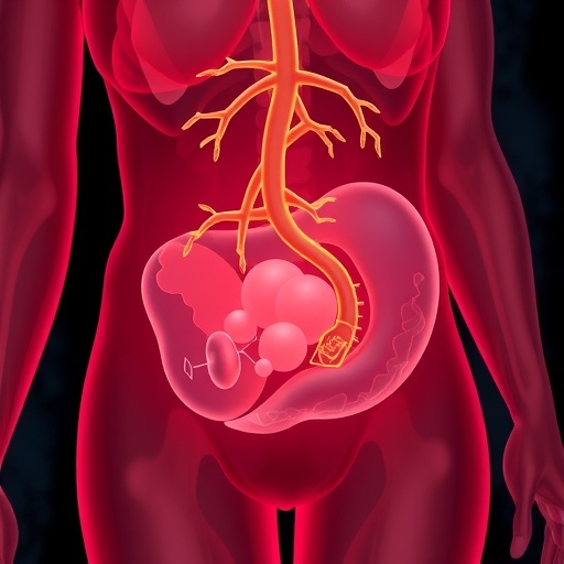
Image Credits: AI Generated
DOI: 10.1038/s41390-026-04957-x
