In an unprecedented exploration of glioblastoma (GBM), one of the most aggressive brain cancers known, a groundbreaking study has delivered profound insights into the tumor microenvironment by combining multiple layers of cutting-edge genomic and spatial technologies. The research, harnessing the immense power of spatial transcriptomics, single-cell RNA sequencing (scRNA-seq), ATAC-seq, and patch sequencing, dissects the complex mosaic of cellular interactions within GBM tissues collected from 100 patients. This comprehensive analysis reveals the intricate cellular communities that orchestrate tumor progression and uncovers novel avenues for therapeutic intervention.
Glioblastoma presents an exceptionally heterogeneous landscape, confounding effective treatment strategies. Traditional methods often overlook the tumor’s spatial and cellular diversity, limiting our understanding of how malignant cells and their microenvironment orchestrate aggressive behavior. By integrating 121 spatial transcriptomic datasets with detailed single-cell profiles, the study captures an unprecedented resolution of the tumor’s cellular architecture. This multi-modal approach enables the mapping of distinct malignant communities and their microenvironmental niches, which sustain and accelerate tumor growth.
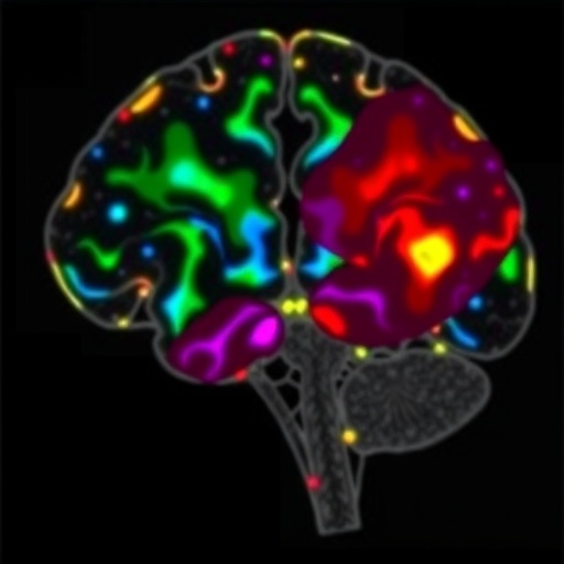
Central to the study’s findings is the identification of four malignant cellular communities consistently observed across patients. These communities form spatially coherent clusters characterized by unique gene expression signatures and cellular behaviors. This discovery shifts the paradigm from viewing GBM as a monolithic mass to understanding it as a complex ecosystem, where cellular communities function dynamically in concert, shaping the tumor’s clinical characteristics.
Among these cellular communities, two distinct subpopulations of mesenchymal-like (MES-like) tumor cells stand out, highlighting the profound heterogeneity even within defined cell lineages. The first subpopulation, termed MES-Hyp, thrives in hypoxic niches and is anatomically interwoven with monocyte-derived brain macrophages. This spatial association hints at a collaborative interplay where immune cells may influence hypoxia-induced tumor evolution and resistance.
The second MES-like subpopulation, termed MES-Ast, exhibits a unique association with vascular elements such as endothelial cells, pericytes, and vascular smooth muscle cells. This cellular neighborhood suggests a role for MES-Ast cells in modulating the tumor vasculature, potentially facilitating nutrient supply and invasive growth. The dichotomy between MES-Hyp and MES-Ast not only underscores the complexity of mesenchymal tumor states but also their functional specialization within the tumor microenvironment.
Beyond the identification of these malignant communities, the study pioneers predictive and experimental validation of cell-type-specific ligand-receptor interactions. These intercellular communications represent molecular conversations that underlie tumor maintenance, immune evasion, and therapeutic resistance. By decoding these signaling networks within each community, the research uncovers previously unrecognized pathways that could be exploited for targeted disruption.
One of the study’s most striking revelations comes from patch sequencing, a technique that combines electrophysiology with single-cell profiling, applied here to tumors in situ. This enabled the discovery that synaptic-like connections between glioma cells and neurons predominantly involve oligodendrocyte-progenitor-like tumor cells (OPC-like). This novel insight suggests that glioma cells not only coexist but intimately interact with neuronal networks, potentially hijacking neural circuitry to support tumor growth and dissemination.
These synaptic interactions open a new frontier in understanding glioma biology, proposing that neural activity and tumor progression are tightly linked—a concept that may revolutionize treatment paradigms by targeting tumor-neuron communication. This insight dovetails with emerging evidence on the role of the nervous system in cancer, moving glioma research into an exciting new neuro-oncology era.
Together, the integrated multi-omic approach delineates a spatial and functional blueprint of the GBM microenvironment, revealing complex cellular ecosystems and dynamic intercellular crosstalk. This spatially resolved molecular atlas provides an invaluable resource for the academic and clinical community, offering maps of cellular states and interactions that drive malignancy and therapeutic resistance.
In clinical terms, these discoveries imply that targeting a single cellular population or signaling pathway might be insufficient, given the tumor’s community-based resilience. Instead, innovative combination therapies disrupting multiple malignant communities and their interactions with the microenvironment might be mandatory to achieve durable responses.
The study also highlights the critical role of tumor-associated macrophages in shaping the hypoxic niche and influencing mesenchymal tumor states, suggesting that modulating immune cell infiltration or function might impair tumor adaptation to harsh microenvironmental conditions and drug resistance.
Vascular-associated MES-Ast cells’ interactions with blood vessel components imply that disrupting tumor-perivascular niches could starve tumors of vital resources and block invasive fronts—potentially enhancing standard chemoradiotherapy efficacy.
Moreover, the identification of ligand-receptor pairs and intercellular communication pathways offers a treasure trove of novel molecular targets. Therapeutic interventions designed to block these molecular dialogs could dismantle the malignant communities’ cooperative networks, rendering the tumor more vulnerable.
This study represents a significant leap forward for personalized neuro-oncology, as the elucidated tumor microenvironmental landscapes differ between patients but maintain overarching cellular community themes. Such knowledge enables stratification of patients based on their tumor’s community composition, enabling precision medicine strategies tailored to disrupt specific pathological interactions.
Technological synergy among spatial transcriptomics, single-cell ATAC-seq to profile chromatin accessibility, and integrative multi-omics bioinformatics set a new standard for tumor microenvironment studies. This multifaceted approach facilitates the construction of a holistic tumor tissue atlas—spatially and functionally annotated at single-cell resolution.
The profound insights into glioblastoma’s cellular ecology gained from this study are expected to galvanize the development of next-generation therapeutic approaches that simultaneously combat tumor heterogeneity and the supportive microenvironment. As the fight against GBM continues, such spatially resolved single-cell analyses may unlock long-elusive vulnerabilities and engender strategies to outsmart this devastating disease.
Taken together, the methodological innovation and biological discoveries presented in this research represent a turning point in glioblastoma research. By unmasking the hidden world of malignant cellular communities and their intimate molecular dialogues, the study lays the groundwork for new therapeutic avenues that can reshape the landscape of brain cancer treatment.
Subject of Research: Glioblastoma tumor microenvironment, spatial transcriptomics, single-cell characterization, intercellular communication, tumor heterogeneity
Article Title: Spatial and single-cell characterization of human glioblastoma tumor microenvironment reveals malignant cellular communities.
Article References:
Lin, J., Chen, C., Li, S. et al. Spatial and single-cell characterization of human glioblastoma tumor microenvironment reveals malignant cellular communities. Nat Neurosci (2026). https://doi.org/10.1038/s41593-026-02265-5
