In recent years, the distressing phenomenon of sea lions beaching along the Southern California coast has garnered increased attention from scientists and conservationists alike. The primary cause of this alarming trend has been traced to toxic algal blooms that sicken these marine mammals, creating an urgent need for more effective medical intervention. Now, a pioneering project led by researchers at the University of Nevada, Las Vegas (UNLV) proposes a transformative approach to wildlife veterinary care by harnessing advances in 3D printing and soft robotics. This technological breakthrough could revolutionize how veterinarians train for and perform life-saving procedures on sea lions, while also laying the groundwork for innovations in human medical devices.
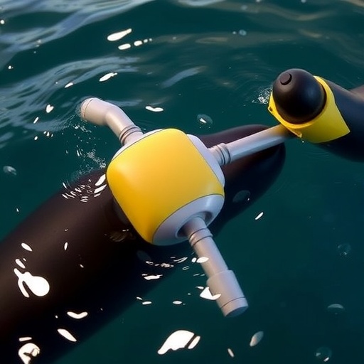
At the core of this research is the creation of hyper-realistic, synthetic models of a California sea lion’s pelvic region. Developed by UNLV’s Active Materials and Smart Living Lab, these models meticulously replicate the detailed anatomy of the pelvic bones and associated soft tissues. What distinguishes these 3D-printed phantoms is their authenticity—not only do they mirror the physical morphology of the biological tissue, but they also emulate tactile qualities such as flexibility, density, and even blood flow characteristics. This fidelity enables veterinary trainees to practice critical blood collection and other invasive procedures in conditions that closely mimic those encountered in live animals.
The genesis of this innovation lies in the harnessing of detailed DICOM data, a standardized file format widely used for storing medical imaging information such as CT and MRI scans. By transforming volumetric imaging data into precise 3D models, researchers extract the intricate vascular and skeletal architecture necessary for faithful reproduction. Advanced software tools convert this data into printable formats that accommodate the nuances of both bone rigidity and soft tissue elasticity. This synergy of medical imaging and additive manufacturing allows the fabrications to serve as functional proxies for living anatomical structures.
Daniel Fisher, a mechanical engineering graduate researcher and lead author of the study, emphasizes the vast potential of this approach. His vision extends beyond improving medical care for marine mammals to encompass future applications involving customized implants and novel surgical techniques for both human and animal patients. The promise lies in the capacity to simulate specific anatomical contexts at an unprecedented level of detail, thereby reducing risks associated with experimental procedures and enhancing clinical outcomes.
Integral to the project’s success is the application of soft robotics principles. Led by Kwang Kim, a distinguished professor and expert in the field, the research exploits smart materials capable of dynamic responses to external stimuli like electrical input or mechanical pressure. These materials mimic the contractility and resilience of natural muscles, enabling the pelvic phantom not only to look anatomically accurate but also to behave in a lifelike manner during manipulation. The result is a training model that provides realistic feedback in tactile sensation and response to needle insertion, closely reproducing the challenges encountered during real interventions.
Beyond training, these synthetic phantoms carry profound implications for surgical planning. Veterinarians and surgeons can now refine their techniques on individualized models tailored to specific animals, gleaned from their own medical imaging data. Such personalization potentially increases procedural efficacy and decreases complications by enabling clinicians to understand precisely how instruments interact with unique anatomical variations. This degree of preparation is especially crucial for endangered or vulnerable species where minimizing harm is imperative.
Further exploring the capabilities of the fabricated phantoms, the UNLV team is investigating how these models perform in aquatic environments, simulating the conditions sea lions naturally inhabit. This research avenue is poised to reveal insights into material durability, fluid dynamics, and anatomical interactions under water, opening new possibilities for the design of implants or assistive devices that function effectively in marine settings. Understanding these dynamics is essential for advancing the field of biomechanical engineering in ways that transcend terrestrial applications.
The broader societal impact of this interdisciplinary research cannot be overstated. The advancements stand at the crossroads of wildlife conservation, robotic engineering, and biomedical innovation, exemplifying how technology can address pressing environmental and health challenges. By improving veterinary care for sea lions, the work contributes directly to marine ecosystem preservation, while also inspiring potential breakthroughs in prosthetics and rehabilitation for humans facing muscular or neurological impairments.
One of the most remarkable aspects of this initiative is its ability to replace reliance on animal carcasses or cadaveric specimens for medical training. Historically, access to authentic biological samples has been limited and fraught with ethical and logistical constraints. The advent of highly accurate synthetic models democratizes educational opportunities, allowing widespread, repeated, and risk-free practice that enhances skill acquisition and procedural confidence among health professionals.
Looking ahead, the research team aspires to incorporate biocompatible materials into their models to develop artificial muscles and implants that could restore lost functions in humans. This translational ambition underscores the versatility of their technology platform, bridging veterinary science and human medicine seamlessly. The pursuit of implanted artificial muscles capable of mimicking natural physiology may redefine therapeutic strategies for conditions previously deemed intractable.
Published in the prestigious journal Scientific Reports, this collaborative study features contributions from UNLV researchers alongside bioengineers affiliated with the U.S. Navy Marine Mammal Program. The support from multiple funding agencies, including the U.S. Army Research Office and the Office of Naval Research, highlights the strategic importance and interdisciplinary appeal of this innovative work. By combining expertise from mechanical engineering, marine biology, and robotics, the authors present a compelling vision for the future of medical training and device development.
In summary, the UNLV-led project demonstrates a sophisticated fusion of 3D printing, soft robotics, and biomedical imaging to create life-like sea lion pelvic models. These tools facilitate improved veterinary training, enable personalized surgical planning, and pave the way for revolutionary implantable devices. Through this blend of science and engineering, the research offers hope for mitigating the devastating impact of toxic algal blooms on marine mammals and, ultimately, enhancing medical outcomes across species. The pioneering spirit embodied in this work not only addresses an urgent ecological crisis but also expands the frontiers of synthetic biology and soft tissue engineering in remarkable ways.
Subject of Research: Animals
Article Title: Scalable DICOM 3D-printed phantoms mimicking marine mammal bone and soft tissue
News Publication Date: January 21, 2025
Web References:
https://www.nature.com/articles/s41598-026-36154-5
References:
Fisher, D., Kim, K., Minaian, N., McClain, A. (2025). Scalable DICOM 3D-printed phantoms mimicking marine mammal bone and soft tissue. Scientific Reports.
Image Credits:
John Domol, University of Nevada, Las Vegas
Keywords
Aquatic animals, Health and medicine, Engineering, Biomaterials
