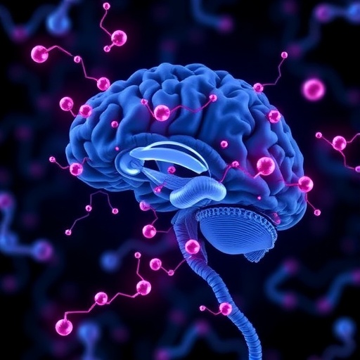
In a compelling breakthrough that could revolutionize our understanding of Parkinson’s disease, researchers have uncovered a critical pathway by which α-synuclein oligomers, derived from cerebrospinal fluid (CSF), propagate pathology in a region-specific manner within the brain. The study, published in npj Parkinson’s Disease, elucidates the perivascular spread of these toxic protein assemblies, offering fresh insights into the mechanisms driving the hallmark neurodegeneration of this devastating disorder. This pioneering research not only sheds light on the elusive transmission routes of α-synuclein aggregates but also opens promising avenues for targeted therapeutic interventions.
For decades, Parkinson’s disease has been chiefly characterized by the abnormal accumulation of α-synuclein protein, forming Lewy bodies that lead to neuronal dysfunction and death. While previous work has established that misfolded α-synuclein can propagate in a prion-like fashion between cells, the precise anatomical and molecular conduits facilitating its spread remained largely speculative. The current investigation by Zhu and colleagues decisively identifies the perivascular spaces as key highways enabling CSF-derived α-synuclein oligomers to infiltrate and seed pathology in susceptible brain regions.
Using advanced biochemical assays combined with innovative imaging techniques, the researchers traced the distribution of α-synuclein oligomers isolated directly from CSF samples. Their analyses revealed a highly selective affinity of these oligomeric species for perivascular niches—zones surrounding blood vessels in the central nervous system that constitute critical interfaces between vascular, glial, and neuronal compartments. These oligomers exploit the perivascular pathways to disseminate, bypassing classic synaptic or neuronal transport routes and selectively targeting regions vulnerable to Parkinsonian degeneration.
The team’s meticulous mapping of α-synuclein deposits demonstrated region-specific accumulation patterns that correlate with symptom onset and progression observed in Parkinson’s disease patients. Crucially, perivascular spread generated distinct foci of α-synuclein pathology in substantia nigra, striatum, and other motor control centers, consistent with clinical phenotypes. These findings elucidate why certain brain areas preferentially undergo neurodegeneration, despite the ubiquitous presence of α-synuclein throughout the nervous system.
Harnessing rodent models engineered to simulate human CSF-derived α-synuclein exposure, the study further validated the perivascular transmission hypothesis. Animals infused with these oligomers displayed progressive motor deficits resembling Parkinsonian symptoms alongside confined but expanding regions of α-synuclein aggregation. Importantly, interventions disrupting perivascular integrity or blocking oligomer interaction with perivascular components significantly attenuated this pathological cascade, underscoring the therapeutic potential of targeting these vascular interfaces.
At a molecular level, the investigation delved deeply into the biochemical properties that empower α-synuclein oligomers to navigate perivascular routes. The oligomers exhibited distinct conformational states and surface-exposed motifs facilitating adhesion to extracellular matrix proteins and pericyte receptors that line vascular boundaries. This selective binding is postulated to be a critical initial step enabling the seeded progression of misfolded α-synuclein aggregates from CSF into brain parenchyma.
Remarkably, the research also highlights the role of the glymphatic system—an emerging brain clearance mechanism involving perivascular fluid flow—in modulating α-synuclein dynamics. Alterations in glymphatic function, as seen in aged or diseased brains, may exacerbate the retention and accumulation of α-synuclein oligomers, thereby accelerating the neurodegenerative process. This link underscores the broader physiological relevance of vascular and fluid homeostasis in neurodegeneration and positions glymphatic modulation as an attractive therapeutic strategy.
The implications of these findings extend beyond unraveling pathogenic α-synuclein spread. They invite a paradigm shift in how scientists conceptualize neurodegenerative disease propagation, emphasizing vascular and perivascular microenvironments as integral players—not merely passive bystanders—in shaping disease trajectory. Moreover, this vascular-centric model may correspondingly inform our understanding of other proteinopathies such as Alzheimer’s disease, which similarly involve perivascular accumulation of pathogenic proteins.
This study’s technological innovations were equally critical for its success. Cutting-edge in vivo imaging of CSF dynamics paired with ultrasensitive α-synuclein oligomer detection refined spatial and temporal resolution of protein dissemination events. Such methodological advances promise to empower future research endeavors aimed at mapping intricate molecular paths of neurodegenerative agents within living brains, facilitating early diagnosis and monitoring of disease progression with unprecedented precision.
While much remains to be explored, Zhu et al.’s work firmly positions perivascular pathways at the forefront of Parkinson’s pathology research. By illuminating how CSF-derived α-synuclein oligomers commandeer perivascular routes to orchestrate region-specific neurodegeneration, this study propels us closer to identifying novel biomarkers and therapeutic targets that could halt or even reverse disease progression. Given the global burden of Parkinson’s and the unrelenting need for effective treatments, such breakthroughs herald new hope for millions worldwide.
In the coming years, further dissecting the cellular and molecular interplay within perivascular niches will be paramount. Understanding how endothelial cells, pericytes, astrocytes, and immune components coordinate to influence α-synuclein trafficking and clearance could unlock strategies to fortify vascular defenses. Moreover, developing small molecules or biologics capable of interrupting oligomer-perivascular interactions or enhancing glymphatic clearance may emerge as viable therapeutic modalities.
This groundbreaking research underscores the power of multidisciplinary collaboration combining neurobiology, vascular physiology, and protein chemistry. It also emphasizes the importance of investigating neurodegeneration through a holistic lens that integrates fluid dynamics, microanatomy, and protein folding pathologies. As we unravel these complexities, the vision of precision medicine tailored to intercept early pathological spread and protect vulnerable brain regions appears increasingly attainable.
The impact of this study extends beyond the laboratory. It invigorates the scientific community’s drive to focus on cerebrovascular and extracellular matrix contributions to neurodegeneration, inspiring new lines of inquiry and funding priorities. Simultaneously, it informs clinicians about the potential significance of vascular health in Parkinson’s disease progression, possibly influencing patient management strategies that blend neurological and cardiovascular care.
Ultimately, the revelation that α-synuclein oligomers exploit perivascular routes to instigate localized Parkinson’s-like pathology marks a significant leap forward in our battle against neurodegenerative disease. Through continued exploration of these vascular pathways and their perturbations, we may soon witness transformative advances in diagnosis, treatment, and prevention that redefine the landscape of Parkinson’s disease and related disorders.
Subject of Research: The regional spread and pathogenic mechanisms of cerebrospinal fluid-derived α-synuclein oligomers in Parkinson’s disease.
Article Title: Perivascular spread of CSF-derived α-synuclein oligomers drives region-specific Parkinson’s-like pathology.
Article References:
Zhu, WX., He, XZ., Meng, JC. et al. Perivascular spread of CSF-derived α-synuclein oligomers drives region-specific Parkinson’s-like pathology.
npj Parkinsons Dis. (2026). https://doi.org/10.1038/s41531-026-01300-3
Image Credits: AI Generated
