Groundbreaking Study Illuminates Normative Growth Trajectories of Fetal Brain Regions with Long-Term Neurodevelopmental Validation
In a pioneering investigation that bridges fetal neuroimaging and early childhood developmental outcomes, researchers have unveiled detailed normative growth trajectories of specific fetal brain regions, confirmed by neurodevelopmental assessments at two years of age. This study offers unprecedented insights into the complex processes of in utero brain maturation and their predictive relationship with postnatal neurological function, potentially transforming prenatal diagnostics and early intervention strategies.
Utilizing state-of-the-art fetal magnetic resonance imaging (MRI) protocols, the research team embarked on a meticulous longitudinal examination of brain structural evolution during critical gestational windows. Unlike previous cross-sectional analyses, this approach allowed for the dynamic mapping of the volumetric and morphological growth patterns of essential brain nuclei and cortical regions from mid-gestation until term. The continuous imaging series generated a high-resolution atlas delineating normative trajectories, surpassing prior attempts limited by smaller cohorts or narrower temporal scopes.
The technical rigor of this study was exemplified by the application of advanced motion correction algorithms and sophisticated brain tissue segmentation techniques, overcoming significant challenges inherent to fetal imaging such as spontaneous fetal movements and variable in utero positioning. Employing novel machine learning models trained on annotated datasets, the authors achieved automated delineation of subcortical structures with exceptional accuracy, enabling reproducible quantification of neuroanatomical volumes essential for growth curve construction.
One of the distinguishing aspects of this research was its integration of fetal brain development data with postnatal neurodevelopmental outcomes, assessed at 24 months using standardized developmental scales encompassing cognitive, motor, language, and social domains. This cross-disciplinary methodology provided a robust framework to validate that the observed prenatal growth trajectories correspond meaningfully with subsequent neurofunctional maturation, thereby underpinning the clinical relevance of normative brain imaging benchmarks.
The longitudinal data illuminated differential growth rates among diverse brain regions, revealing nuanced patterns such as the accelerated volumetric expansion of the hippocampus and cerebellum relative to cortical areas during the late second and third trimesters. These findings underscore the heterochronous nature of fetal brain maturation and emphasize the importance of region-specific normative data to discern typical from aberrant developmental courses.
Delving deeper into neurodevelopmental implications, the study identified specific prenatal growth trajectories that significantly correlate with improved outcomes in executive function and language acquisition at toddlerhood. This linkage supports emerging hypotheses that fetal neuroanatomical development kinetics serve as early biomarkers for cognitive capacities, highlighting critical windows during which deviations might forecast developmental delays or disorders.
The comprehensive normative dataset generated by this study holds profound translational potential. Clinicians can now benchmark individual fetal MRI scans against validated growth curves, enhancing the detection of in utero brain pathologies such as microcephaly, ventriculomegaly, or hypoplasia within the context of population-based standards. Early identification of deviations could prompt timely interventions, optimizing therapeutic windows during prenatal or early postnatal periods.
From a technical standpoint, the study innovated in the harmonization of multi-center MRI acquisition protocols, leveraging standardized imaging parameters and calibration phantoms to ensure data comparability. This pioneering effort addresses the long-standing obstacle of variability in fetal MRI studies, paving the way for large-scale multi-institutional collaborations and meta-analyses that can refine and extend normative brain growth paradigms.
Moreover, the research team explored the influence of maternal-fetal environmental factors including maternal age, nutrition, and gestational health conditions on fetal brain growth patterns. Although subtle, discernable modulations in certain brain regions were detected, suggesting that these variables may impact neurodevelopment trajectories. Future investigations may unravel specific mechanisms mediating these associations, contributing to more personalized prenatal care strategies.
Significantly, this study adopted a machine learning predictive modeling approach, using prenatal brain morphometric data to estimate neurodevelopmental scores at two years with impressive accuracy. Integrating neuroimaging features with demographic and clinical covariates, these models represent a leap towards presymptomatic identification of infants at risk for neurodevelopmental disorders, a paradigm shift in fetal medicine.
Ethical considerations were rigorously addressed throughout the study, ensuring voluntary informed consent and rigor in data anonymization. The transparency of the methodology and data accessibility initiatives promote reproducibility and collective advancement in fetal neuroscience, fostering an open scientific environment vital for accelerating progress.
This landmark investigation not only enhances our understanding of normative fetal brain maturation but also sets a gold standard for future neurodevelopmental research spanning prenatal to early childhood stages. Its publication in a prestigious scientific journal affirms the critical importance of merging cutting-edge imaging, computational analysis, and developmental psychology to unravel the foundations of human brain development.
As fetal MRI technology continues to evolve, integrating functional imaging modalities such as fetal functional MRI and spectroscopy could further elucidate the intricate relationship between structural growth and emerging cerebral functions. The comprehensive normative framework established here will serve as a cornerstone for such multidimensional studies, advancing early diagnosis and intervention for neurodevelopmental disorders.
The implications of this work extend beyond clinical practice to inform public health policies focused on maternal and child health. By elucidating normative brain growth parameters, policymakers can better assess population-level fetal well-being and prioritize resources for prenatal screening programs targeting high-risk pregnancies.
In summary, this study by Wyburd, Kennedy, Fernandes, and colleagues epitomizes the confluence of technological innovation, methodological precision, and clinical relevance. Their contribution charts new territories in fetal brain research, reinforcing the concept that the prenatal period is a critical foundation stage for lifelong neurological health and cognitive potential. The future of fetal neuroimaging stands poised on the threshold of transformative discoveries, driven by studies such as this that marry statistical modeling with biological insight.
Subject of Research: Normative growth trajectories of fetal brain regions and their validation through neurodevelopmental domain maturation at two years of age.
Article Title: Normative growth trajectories of fetal brain regions validated by satisfactory maturation of neurodevelopmental domains at 2 years of age.
Article References:
Wyburd, M.K., Kennedy, S.H., Fernandes, M. et al. Normative growth trajectories of fetal brain regions validated by satisfactory maturation of neurodevelopmental domains at 2 years of age. Nat Commun (2026). https://doi.org/10.1038/s41467-026-69657-w
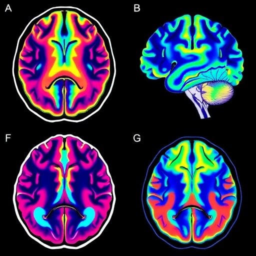
Image Credits: AI Generated
