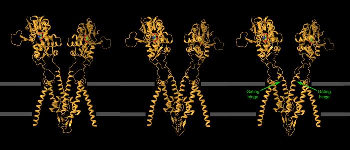
NEW YORK, NY–New groundbreaking images of one of the brain’s fastest-acting proteins are providing critical clues that may lead to the development of targeted therapies to treat epilepsy and other brain disorders.

Credit: Columbia University Irving Medical Center
NEW YORK, NY–New groundbreaking images of one of the brain’s fastest-acting proteins are providing critical clues that may lead to the development of targeted therapies to treat epilepsy and other brain disorders.
The lightning-fast moves of the brain’s kainate receptor are indispensable for neuron-to-neuron communication but create a quandary for structural biologists trying to capture images of the receptor in action.
When called into action, a kainate receptor embedded in the surface of a neuron opens its ion channel, then slams the channel shut within milliseconds.
“That’s the key problem for structural biologists: You have to freeze these molecules moments before the channel closes,” says Alexander Sobolevsky, associate professor of biochemistry & molecular biophysics at Columbia University Vagelos College of Physicians and Surgeons, who led the team that obtained the new images.
“But it takes about 30 seconds to freeze a molecule, and that’s just too slow.”
It’s not that structural biologists—scientists who obtain images and build models of life’s molecules—haven’t tried.
Dysfunction in the kainate receptor is involved in epilepsy and has been implicated in many other brain disorders, including depression, anxiety, and autism. It’s a desirable target for drug developers, but only a few approved drugs safely target the receptor.
“Everyone is trying to get images of the receptor in action so we can understand how it works and design drugs to control it,” Sobolevsky says.
Success with two innovations
“Capturing images of the active receptor was a long-standing scientific problem that we were trying to solve,” says Shanti Pal Gangwar, an associate research scientist in Sobolevsky lab. The amount of time that the channel remains open during activation, a few milliseconds, is too short to perform conventional freezing of the receptor for cryo-electron microscopy (cryo-EM), the structural biology technique used by Sobolevsky’s lab to image life’s molecules.
To obtain images of the receptor in action, the Sobolevsky team developed two different tricks that slow channel closure and speed up the freezing process.
First, the team looked for molecules that stick to the receptor and keep it open. They found two such molecules—BPAM and lectin Concanavalin A. The small drug-like molecule BPAM prolonged the channel opening by a few hundred milliseconds, but the delay was still insufficient to capture the channel in an open state. Only after addition of the lectin, a protein that binds to sugars decorating glycoproteins, did the channel remain open for several seconds.
Electrophysiological recordings were key to figuring out the timing. “With these recordings, I found that BPAM or lectin alone was incapable of maintaining the channel open long enough,” says Maria Yelshanskaya, an associate research scientist in Sobolevsky lab and an expert in electrophysiology. “When I saw the synergistic effect of BPAM and lectin together, it was like a Eureka moment for us.”
Several seconds was still too fast for conventional cryo-EM freezing, which takes about 30 seconds to plunge the sample into liquid ethane and ready it for imaging. So Kirill Nadezhdin, a postdoctoral research scientist in the Sobolevsky lab, designed a robotic plunger to dunk the sample in less than 3 seconds, trapping the receptors (bound to BPAM and lectin) in an open state.
With the robotic plunger and the molecular stabilizers, Sobolevsky’s team captured images of the kainate receptor in a variety of open configurations.
“Both innovations were necessary,” Sobolevsky says. “We wouldn’t have been able to capture the receptor’s open states with just one or the other.”
Images provide clues for new drugs
The images of the open kainate receptor provide critical information for drug developers.
Some drugs, ion channel blockers, act like corks in wine bottles and plug open channels. “You need the structure of an open channel in order to create a drug that is a perfect, atom-to-atom fit,” Sobolevsky says.
The images also show how perampanel, an epilepsy drug that targets kainate receptors, could be optimized to target specific versions of kainate receptors and provide more precise medicines for patients. Already, collaborators are using Sobolevsky’s lab images to explore with computer modeling possible changes to the drug.
The methods developed by Sobolevsky team should help other investigators obtain more images of open kainate receptors and other types of molecules that work quickly.
“The more images we have, the better the models will be, and hopefully the better our drugs will be,” Sobolevsky says.
Additional information
“Kainate receptor channel opening and gating mechanism,” was published online May 22 and appears in the June 20 print issue of Nature.
All authors (from Columbia unless noted): Shanti Pal Gangwar, Maria V. Yelshanskaya, Kirill D. Nadezhdin, Laura Y. Yen, Thomas P. Newton, Muhammed Aktolun (Carnegie Mellon University), Maria G. Kurnikova (Carnegie Mellon University), and Alexander I. Sobolevsky.
This research was supported by the NIH (grants U24GM129547, U24GM129539, U24GM129541, NS083660, NS107253, AR078814, and CA206573), the Simons Foundation (SF349247), and NY State Assembly Majority. Some of this work was performed at the Columbia University Cryo-Electron Microscopy Center, the Pacific Northwest Center for Cryo-EM at Oregon Health & Science University, the National Center for CryoEM Access and Training (NCCAT), the Simons Electron Microscopy Center at the New York Structural Biology Center, and the Stanford-SLAC Cryo-EM Center.
The authors report no competing interests.
###
Columbia University Irving Medical Center
Columbia University Irving Medical Center (CUIMC) is a clinical, research, and educational campus located in New York City, and is one of the oldest academic medical centers in the United States. CUIMC is home to four professional colleges and schools (Vagelos College of Physicians and Surgeons, Mailman School of Public Health, College of Dental Medicine, and School of Nursing) that are global leaders in their fields. CUIMC is committed to providing inclusive and equitable health and medical education, scientific research, and patient care, and working together with our local upper Manhattan community—one of New York City’s most diverse neighborhoods. For more information, please visit cuimc.columbia.edu.
Journal
Nature
Article Title
Kainate receptor channel opening and gating mechanism
Article Publication Date
22-May-2024
COI Statement
No disclosures.


