In a groundbreaking advancement merging artificial intelligence with oncology, a recent study unveils a multimodal AI system capable of accurately predicting metastasis in cutaneous melanoma by integrating diverse data streams representing the tumor microenvironment. This innovative approach marks a significant leap forward in the personalized prognosis and management of melanoma, a notoriously aggressive skin cancer often complicated by unpredictable metastatic spread. The research, led by Andrew, T.W., Combalia, M., and Hernandez, C., and published in Nature Communications, harnesses sophisticated computational models that analyze and synthesize histopathological features alongside molecular and spatial data to achieve unprecedented predictive accuracy.
Cutaneous melanoma is a formidable clinical challenge due to its propensity for rapid progression and potential to metastasize to distant organs, which dramatically worsens patient outcomes. Traditional prognostic models rely heavily on tumor thickness, ulceration status, and nodal involvement, but these factors alone often fail to capture the complex biological interplay dictating metastatic potential. Recognizing this gap, the research team focused on the tumor microenvironment (TME)—the dynamic ecosystem surrounding malignant cells composed of immune components, stromal cells, and extracellular matrix elements—as a vital source of prognostic information. The TME not only influences tumor growth but also modulates immune surveillance and therapy responsiveness, making it a fertile ground for analysis.
At the heart of the study is a sophisticated AI framework that integrates multiple data modalities—digital pathology images, molecular profiling, and spatial cellular interactions—to decode the multifaceted nature of melanoma progression. The team employed deep learning techniques to process whole-slide histopathological images, extracting features that reveal architectural nuances of tumor and surrounding tissues. Concurrently, molecular data provided a high-resolution map of gene expression patterns and protein markers, while spatial analyses captured the distribution and interaction networks of immune and stromal cell populations within the tumor niche. This multimodal fusion allows the AI to synthesize diverse biological signals into a cohesive predictive model.
A key technical innovation in this work is the utilization of convolutional neural networks (CNNs) tailored for histopathological image analysis, combined with graph neural networks (GNNs) that effectively model cellular interactions within the tissue architecture. The CNNs parse complex visual patterns that elude traditional pathology, identifying subtle cues associated with aggressive behavior. Meanwhile, GNNs map spatial proximity and communication pathways among cells, elucidating how immune cells and tumor cells influence each other’s behavior spatially. By combining these architectures, the AI system gains a holistic understanding of tumor biology that reflects the true complexity of the TME.
To train and validate the model, the researchers assembled an extensive dataset comprising digital pathology slides and corresponding molecular data from hundreds of melanoma patients, including long-term follow-up information on metastatic outcomes. Cross-validation and independent test cohorts were employed to rigorously assess the model’s performance. The AI system demonstrated superior accuracy and prognostic power compared to current clinical staging systems, with particularly strong predictive ability in early-stage melanoma cases where traditional risk stratification is challenging. This capacity to preemptively identify high-risk patients could inform more tailored surveillance and therapeutic interventions.
One of the most notable aspects of this multimodal AI model is its interpretability—a critical feature for clinical adoption. By implementing attention mechanisms within the neural networks, the system highlights specific histopathological regions, molecular markers, and cellular interactions that most heavily contribute to its metastasis predictions. This feature not only provides transparency but also offers valuable biological insights, revealing previously underappreciated TME components that drive melanoma dissemination. Such discoveries open new avenues for therapeutic targeting and biomarker development.
The integration of spatial profiling data into the AI pipeline constitutes a major stride in oncology research. The tumor microenvironment is not static; it is shaped by the spatial orchestration of immune cells, fibroblasts, and endothelial cells, which collectively dictate tumor evolution and resistance mechanisms. By capturing these spatial dynamics, the AI system can discern micro-anatomical niches that either suppress or facilitate tumor spread. This spatially aware approach represents a departure from prior models that treated tumors as homogeneous entities and underscores the importance of cellular geography in cancer progression.
From a computational standpoint, the study tackles several challenges inherent in multimodal data integration, including heterogeneity in data types, scaling issues, and alignment of spatial coordinates across imaging and molecular datasets. The researchers implemented advanced data normalization techniques and cross-modal embedding strategies that create a unified latent space, enabling seamless communication between diverse data formats. This methodological rigor ensures that insights drawn from one modality complement and enhance findings from others, leading to a more robust predictive framework.
Beyond mere prediction, the AI framework serves as a discovery engine unveiling the complex biology underpinning melanoma metastasis. Patterns uncovered by the model suggest that immune evasion strategies, extracellular matrix remodeling, and localized hypoxic zones within the TME are critical determinants of metastatic spread. Such mechanistic insights could redefine how clinicians assess tumor aggressiveness and tailor treatments, shifting the paradigm toward truly precision oncology.
Importantly, the study also addresses the translational potential and challenges of applying this AI tool in routine clinical settings. The authors discuss the feasibility of integrating the multimodal AI system with existing diagnostic workflows, emphasizing the need for standardized data acquisition protocols, computational infrastructure, and clinician training. By providing a clear pathway for implementation, the research bridges the gap between technological innovation and practical healthcare impact.
This work resonates within the broader context of AI-driven medicine where combining multimodal datasets holds promise for unraveling complex diseases. Melanoma, with its rich pathological heterogeneity and diverse molecular landscape, serves as a compelling testbed demonstrating how artificial intelligence can transform diagnostic precision and therapeutic decision-making. The success of this model heralds a new era where digital pathology, spatial biology, and computational analytics converge to tackle enduring clinical challenges.
Furthermore, the implications extend beyond melanoma, as the AI methodology developed here is adaptable to other malignancies where the tumor microenvironment plays a pivotal prognostic role. Cancers characterized by intricate stromal-immune interactions such as breast, lung, and colorectal carcinomas could benefit from similar multimodal analytical approaches. This versatility underscores the transformative potential of AI in oncological research and clinical practice.
As the AI system continues to evolve, future work will likely incorporate additional data layers such as single-cell sequencing, metabolomics, and longitudinal imaging to further enhance predictive accuracy and biological insight. Continuous refinement and external validation across diverse populations will be crucial to ensure generalizability and equity in cancer care. The integration of patient-specific clinical variables alongside molecular and spatial data may also provide even more personalized risk assessments.
In summary, the pioneering work presented establishes a powerful AI platform that not only anticipates metastatic risk in cutaneous melanoma with unprecedented accuracy but also deciphers the intricate tumor microenvironmental landscape driving disease progression. This fusion of multimodal data modalities through advanced machine learning propels both scientific understanding and clinical capability forward, opening the door to more effective, individualized cancer management strategies. The intersection of technology and biology showcased here exemplifies the future of precision oncology—where intelligent systems illuminate the path toward better patient outcomes.
Subject of Research: Prediction of metastasis in cutaneous melanoma through integration of multimodal AI and tumor microenvironment data.
Article Title: Multimodal AI and tumour microenvironment integration predicts metastasis in cutaneous melanoma.
Article References:
Andrew, T.W., Combalia, M., Hernandez, C. et al. Multimodal AI and tumour microenvironment integration predicts metastasis in cutaneous melanoma. Nat Commun 16, 10095 (2025). https://doi.org/10.1038/s41467-025-65051-0
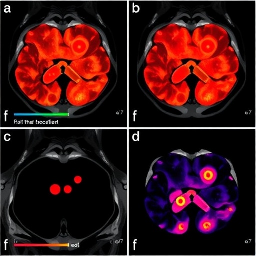
Image Credits: AI Generated
