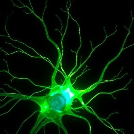
In a groundbreaking study poised to reshape our understanding of nerve repair, researchers have unveiled the intricate molecular choreography behind axon regeneration in the adult central nervous system (CNS). Unlike the peripheral nervous system and developing neurons during embryogenesis, adult CNS neurons notoriously fail to regenerate after injury, leaving the brain and spinal cord vulnerable to permanent damage. This longstanding enigma has perplexed neuroscientists for decades, but the latest research sheds light on the mechanisms that can unlock regeneration and potentially pave new paths for treating debilitating CNS injuries.
At the heart of this discovery lies the drug epothilone B, a molecule previously known to stabilize microtubules, the dynamic structural filaments that compose the cellular cytoskeleton. Microtubules play a vital role in maintaining cell shape, intracellular transport, and crucially, the extension of neuronal processes. Employing a sophisticated combination of in situ cryo-electron tomography and cryo-electron microscopy, the research team has mimicked axonal damage within live neuronal tissue, capturing snapshots of the cellular response at near-atomic resolution. These unprecedented images reveal how stabilized microtubules extend beyond injury sites, generating mechanical forces that drive membrane expansion and initiate axonal regeneration.
The in situ cryo-electron microscopy data, resolved to 3.19 angstroms, uncovered the binding of epothilone B deep within the architecture of microtubules precisely at the regenerating front of injured axons. This binding appears to enhance microtubule stability, allowing polymerization processes to persist despite cellular trauma. Significantly, the researchers observed active delivery of tubulin clusters— the building blocks of microtubules— to the injury site, where these subunits incorporate into growing microtubule “shoots.” These shoots act not only as structural scaffolds but also as conduits for transporting vesicles and endoplasmic reticulum, essential components for membrane repair and axonal growth.
This study overturns previous assumptions that adult CNS neurons lack sufficient intrinsic capacity for regeneration. Instead, it reveals a previously unappreciated resilience, showing neurons’ remarkable ability to adjust to strain induced by epothilone B. By generating localized membrane tension through microtubule extension, axons enter a “regeneration mode,” activating cellular pathways aligned with repair and growth. This paradigm shift suggests that rather than a global inability to regenerate, adult CNS neurons may require precise molecular and mechanical cues that can be pharmacologically induced.
The implications of these findings are profound, opening avenues for novel therapeutic interventions against CNS injuries such as spinal cord damage, stroke, and neurodegenerative diseases. The detailed structural insight provided by cryo-electron microscopy serves as a blueprint for designing drugs that mimic or enhance the microtubule-stabilizing effects of epothilone B, potentially overcoming the inhibitory environment typically encountered by regenerating axons in the CNS milieu. Moreover, understanding how membrane tension and microtubule dynamics interplay may inform biomaterial engineering efforts to create scaffolds that support neuronal repair.
Importantly, the study highlights the cellular logistics of regeneration, emphasizing how microtubule shoots serve as highway systems for intracellular trafficking. The vesicles and endoplasmic reticulum ferry lipids, proteins, and signaling molecules necessary to rebuild the axonal membrane and restore functional connectivity. This integrated approach unites the mechanical, structural, and biochemical facets of neuronal repair, painting a holistic picture of CNS regeneration.
Previously, the role of microtubules in axon regeneration was appreciated mostly from static or ex vivo studies. The present work’s in situ methodology maintains the native cellular environment, providing dynamic snapshots revealing how microtubules polymerize and how epothilone B interacts with the lattice in real time. Such high-fidelity imaging techniques represent a powerful new frontier in neuroscientific research, capable of resolving sub-cellular processes with atomic precision under physiologically relevant conditions.
Beyond therapeutic prospects, these findings stimulate fundamental questions about neuronal plasticity and resilience throughout adulthood. They challenge dogmas around the fixed nature of CNS neurons and inspire exploration of other molecular agents or mechanical stimuli that could similarly unleash regenerative programs. This could ultimately revolutionize how we approach brain and spinal cord injuries, moving from symptomatic care to true biological repair.
The work also underscores the value of interdisciplinary collaboration, combining advances in structural biology, neuropharmacology, and advanced imaging modalities. It sets a benchmark for future studies aiming to decipher the molecular underpinnings of complex cellular behaviors within intact tissues, bridging the gap between molecular mechanisms and organismal outcomes.
Epothilone B’s role as a microtubule-stabilizing agent was known in oncology and neurobiology, but this study positions it as a key molecule unlocking previously inaccessible regenerative capabilities of CNS neurons. By stabilizing microtubules and facilitating the transport of critical cellular components, it functions as both a mechanical and biochemical inducer of repair, providing a dual stimulus essential for overcoming the inhibitory landscape of neural injury.
Ultimately, this research catapults us closer to developing effective strategies for restoring lost neuronal connections and function after CNS trauma. The detailed mechanistic insights offer a compelling rationale for clinical exploration of epothilone B derivatives or related compounds in CNS regeneration therapies. The capacity of neurons to enter a regenerative state through targeted molecular intervention marks a hopeful milestone in neuroscience.
As our ability to visualize and manipulate the subcellular landscape advances, so too does our potential to transform devastating CNS injuries into conditions amenable to recovery. The convergence of molecular precision, mechanical understanding, and therapeutic innovation embodied in this study heralds a new era in neuroregenerative medicine, where the once-impossible dream of CNS axon repair inches toward reality.
Subject of Research: Central nervous system axon regeneration and microtubule stabilization.
Article Title: In situ structural mechanism of epothilone-B-induced CNS axon regeneration.
Article References:
Bodakuntla, S., Taira, K., Yamada, Y. et al. In situ structural mechanism of epothilone-B-induced CNS axon regeneration. Nature (2025). https://doi.org/10.1038/s41586-025-09654-z
