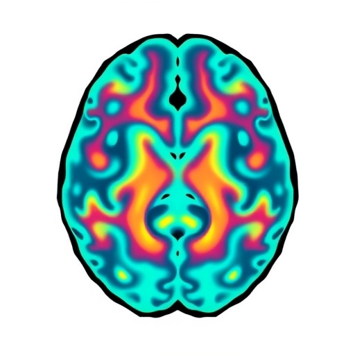
In a remarkable stride toward enhancing pediatric brain tumor diagnostics, researchers at Capital Medical University have developed a novel artificial intelligence (AI) model capable of detecting medulloblastoma subtypes and associated genetic risk factors from magnetic resonance imaging (MRI) scans. Medulloblastoma stands as the most prevalent malignant brain tumor in children, yet its clinical course varies dramatically depending on the tumor’s molecular subtype. Traditional approaches to classify these subtypes require invasive biopsy procedures, which not only pose potential risks to patients but also introduce delays in critical treatment decisions. This breakthrough AI tool, detailed in a forthcoming publication in the Chinese Neurosurgical Journal, presents a promising avenue to accelerate and refine medulloblastoma diagnostics through non-invasive imaging techniques.
The study was spearheaded by Dr. Yanong Li, an expert in radiation oncology at Beijing Tiantan Hospital, part of Capital Medical University, China. Central to the research was the design and training of a convolutional neural network, dubbed MB-CNN, which was exposed to a substantial dataset of MRI scans from 449 pediatric patients treated between 2015 and 2023. The model was meticulously trained to differentiate four principal molecular subgroups of medulloblastoma: the wingless (WNT), sonic hedgehog (SHH), Group 3, and Group 4 categories. Each subtype is characterized by distinct genetic profiles and clinical prognoses, necessitating tailored therapeutic approaches. The AI demonstrated strong proficiency, accurately classifying tumor subtypes in approximately 78% of cases.
What sets the MB-CNN apart is its dual-function capacity—not only can it classify tumor subtypes, but it can also predict specific genetic alterations linked to treatment outcomes and prognosis. These include mutations of the TP53 gene within SHH tumors, amplification of the MYC oncogene in Group 3 tumors, and the deletion of chromosome 11 in Group 4 tumors. The ability to infer such precise genetic details directly from MRI scans represents a major advancement since these genetic tests traditionally require complex and time-consuming laboratory assays on tumor tissue. Impressively, the AI model predicted these genetic alterations with high accuracy, achieving 91% sensitivity for TP53 mutations, 84% for MYC amplifications, and 87% for chromosomal deletions.
Dr. Li emphasized the clinical impact of these capabilities, stating, “Our objective is to provide clinicians with a rapid, less invasive tool that elucidates a patient’s tumor molecular subgroup and relevant genetic risks, thereby guiding more informed and timely treatment decisions.” The implications are profound: by reducing dependence on invasive biopsy procedures and genetic testing turnaround times, this AI-driven diagnostic process could streamline patient management, especially in resource-limited settings where advanced genetic sequencing technologies are not readily accessible.
To benchmark their model’s performance, the team compared MB-CNN against a conventional classification method that relied solely on clinical and radiological data. The traditional approach yielded an accuracy of roughly 59%, underscoring the limitations of existing diagnostic paradigms. Integration of the AI predictions with clinical data into a hybrid model further enhanced classification accuracy, pushing it to an impressive 82.2%. This synergy underscores the potential of combining AI imaging analytics with traditional diagnostic information to optimize medulloblastoma subgroup identification.
Despite these encouraging results, the researchers caution that the study’s retrospective design and the variability introduced by differences in MRI scanners across two institutions might influence the robustness of model performance. To validate and generalize these findings, expansive prospective, multicenter studies are imperative. Such investigations would assess the model’s clinical utility across diverse healthcare environments and heterogeneous patient populations, ensuring its reliability before broad clinical adoption.
From a technical standpoint, MB-CNN represents a sophisticated deep learning framework that harnesses convolutional neural networks to detect subtle image patterns imperceptible to human radiologists. The neural network architecture extracts hierarchical features from MRI scans, learning nuanced morphological and textural cues corresponding to distinct medulloblastoma molecular subtypes and their associated genetic signatures. This level of precision in noninvasive diagnostics could revolutionize how clinicians stratify risk and personalize therapy for pediatric brain tumor patients.
Looking forward, Dr. Li envisions the integration of AI-based imaging models into routine molecular diagnostics workflows. “This research is a critical step toward embedding AI within precision oncology,” he remarked. “While it will not replace traditional genetic assays, such technology can augment decision-making processes by providing swift molecular insights, facilitating earlier interventions, and potentially improving clinical outcomes.” Furthermore, AI-enabled imaging analytics might serve as a triage tool to prioritize urgent cases or identify candidates for specific targeted therapies.
Capital Medical University’s rigorous funding support from top-tier national and municipal science foundations underscores the strategic importance of this research. Backing from bodies including the National Natural Science Foundation of China and the Beijing Nova Star Program reflects growing recognition of AI’s transformative potential within medical diagnostics. Continued financial investments will be pivotal for advancing AI applications from experimental models to clinically validated tools that can be seamlessly adopted worldwide.
Beijing Tiantan Hospital remains at the forefront of neurological research in China, fostering innovations that blend clinical expertise with cutting-edge technology. The development of MB-CNN epitomizes the institution’s commitment to pioneering solutions addressing complex neurological disorders like medulloblastoma, ultimately striving to enhance patient care through multidisciplinary collaboration and scientific discovery.
In conclusion, this pioneering AI model leverages deep learning to noninvasively identify medulloblastoma subtypes and prognostic genetic markers from routine MRI scans, achieving accuracy far superior to conventional approaches. Its capacity to streamline molecular risk assessment promises to reshape pediatric neuro-oncology diagnostics by accelerating personalized treatment planning, minimizing invasive procedures, and expanding access to molecular insights. As further validation studies ensue, MB-CNN may emerge as a vital tool in enhancing outcomes for children afflicted by this challenging brain tumor.
Subject of Research: People
Article Title: Exploring deep learning and hybrid approaches in molecular subgrouping and prognostic-related genetic signatures of medulloblastoma
News Publication Date: 15-Sep-2025
Web References: http://dx.doi.org/10.1186/s41016-025-00405-7
Image Credits: Dr. Yanong Li from Capital Medical University, Japan
Keywords: Health and medicine, Clinical medicine, Health care, Medical diagnosis, Medical tests, Cancer risk, Cancer screening, Cancer patients, Artificial intelligence, Medical technology, Biomedical engineering, Bioengineering
