In a groundbreaking study published in 2025, researchers have embarked on a pioneering exploration of the potential for multimodal Magnetic Resonance Imaging (MRI) techniques to quantify brain lipids in a murine model. This research is critical given the significant role lipids play in various neurological disorders, including Alzheimer’s disease and other neurodegenerative conditions. The study, conducted at a robust magnetic field strength of 9.4 Tesla, marks a notable advancement in neuroimaging technology, highlighting its capability to provide finer resolutions and insights into the brain’s lipid composition compared to standard imaging methods.
The study was designed to traverse the complexities of lipid biochemistry in the neurological system, specifically focusing on how alterations in lipid profiles can signal pathological changes. Researchers, led by Khokhar, Swain, and Soni, utilized advanced MRI protocols that combine multiple imaging modalities to discern lipid concentrations with unprecedented precision. This multifaceted approach underscores the importance of utilizing a comprehensive analysis framework that can capture the dynamic nature of lipid metabolism in the brain.
While conventional imaging techniques have provided valuable insights into brain structure and functionality, they often fall short in differentiating lipid species and their specific contributions to neurological health. The integration of multiple MRI modalities in this research not only enhances spatial resolution but also improves the specificity of lipid detection. This nuanced understanding is vital for researchers and clinicians alike, as it bridges the gap between structural abnormalities in the brain and their biochemical correlates.
At the heart of the methodology employed in this study lays the use of high-resolution proton magnetic resonance spectroscopy alongside diffusion-weighted imaging and chemical shift imaging. This combination allows for a detailed examination of lipid content and distribution across different regions of the brain. The ability to visualize and measure brain lipids in vivo opens new avenues for the study of lipid-related disorders, as it allows researchers to assess lipid profiles without the need for invasive procedures.
Moreover, the findings from this study have the potential to revolutionize the way we approach the diagnosis and monitoring of neurodegenerative diseases. By establishing a connection between lipid profiles and disease states, physicians may soon have the tools they need to develop more targeted therapeutic strategies. It is anticipated that these advancements could also facilitate the early detection of conditions like Alzheimer’s, where early intervention is key to slowing disease progression.
In addition to the implications for diagnosing and understanding neurodegenerative disorders, this research highlights the broader significance of lipid metabolism in brain health. The brain is a highly lipid-rich organ, and its lipid composition is critical for maintaining cellular integrity, supporting neurofunction, and modulating signaling pathways. Understanding the intricate relationship between lipid profiles and brain function can help elucidate mechanisms underlying various psychiatric disorders as well.
This study also brings to light important considerations regarding the animal models used in this research. Mice, which are often used in biomedical research, provide valuable insights into human disease due to their genetic, biological, and behavioral similarities to humans. However, translating findings from murine models to human applications remains a challenge that researchers continually seek to address. The multimodal MRI approach lays the groundwork for future research that could be adapted to human studies, bridging the gap between animal and clinical research.
Beyond the immediate implications for neuroscientific research, this work also emphasizes the utility of advanced imaging technologies in basic science and clinical practice. Continual advancements in MRI technology, such as the capabilities offered by 9.4T imaging, provide researchers with increasingly powerful tools to investigate the brain and its functions. This will pave the way for improved diagnostic techniques and therapeutic approaches that rely on a more sophisticated understanding of lipid dynamics in the central nervous system.
It is also worth noting that as the field of imaging continues to evolve, ongoing research like this will likely inspire collaborations between neuroscientists, radiologists, and bioengineers. Such interdisciplinary partnerships will be crucial for translating these advanced imaging techniques into routine clinical practice, ensuring that the benefits of novel research are accessible to patients and healthcare providers alike.
As the study’s authors articulated, the integration of multimodal MRI techniques holds promise not only in academic research settings but also in the broader context of public health. As we begin to understand the significant influence of brain lipids on overall health and disease, the potential for early intervention through enhanced imaging and lipid profiling could lead to significant improvements in outcomes for individuals suffering from neurodegenerative diseases.
In conclusion, the work conducted by Khokhar, Swain, Soni, and colleagues marks an important step forward in brain imaging research. By leveraging advanced multimodal MRI techniques to quantify brain lipids at 9.4T, this study sets a new standard for how we approach the study of lipid metabolism in the brain. While more research is necessary to fully elucidate the clinical applications of these findings, the study undoubtedly advances our understanding of the intricate relationship between brain health and lipid dynamics.
This research stands as a testament to the power of innovation in science and the importance of continual exploration in understanding complex biological systems. As researchers, we remain hopeful that such studies will fuel further investigations into the intricate workings of the brain, ultimately leading to transformative improvements in the treatment and prevention of neurodegenerative diseases.
Through this groundbreaking work, the scientific community is encouraged to continue pushing the frontiers of research, exploring the depths of human health, and leveraging technological advancements to unravel the mysteries of the brain.
Subject of Research: Brain lipid quantification using multimodal MR imaging.
Article Title: Multimodal MR imaging for quantification of brain lipid in mice at 9.4T.
Article References:
Khokhar, S.K., Swain, A., Soni, N.D. et al. Multimodal MR imaging for quantification of brain lipid in mice at 9.4T.
J Transl Med (2025). https://doi.org/10.1186/s12967-025-07476-1
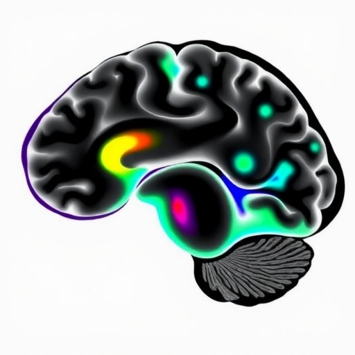
Image Credits: AI Generated
DOI: 10.1186/s12967-025-07476-1
Keywords: Multimodal MRI, brain lipids, neuroimaging, neurodegenerative diseases, lipid profiling, in vivo imaging, neurobiology.
