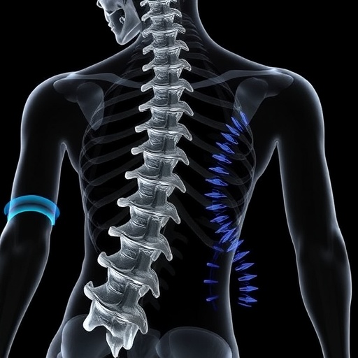
In a groundbreaking study, researchers from various fields have collaborated to explore the innovative application of musculoskeletal ultrasound technology in understanding lumbar spine movements through advanced three-dimensional kinematics visualization. The research, published in the Journal of Medical and Biological Engineering, investigates the energy potential that musculoskeletal ultrasound holds for non-invasive analysis of spinal motion. With its ability to provide real-time imaging, this technology may change how clinicians diagnose and treat lumbar spine disorders.
The study offers an impressive combination of clinical insight and technological development, showcasing how ultrasound—a tool traditionally used for soft tissue evaluation—can be adapted for the analysis of complex musculoskeletal interactions. Researchers Effatparvar, St-Pierre, and Lavoie, among others, dissect the mechanics behind lumbar spine movement through their in vitro analyses, laying the groundwork for future applications in orthopedic assessments. Their findings attempt to bridge the gap between traditional imaging techniques and modern physical analysis methods.
The lumbar spine, composed of vertebrae, discs, and surrounding musculature, plays a critical role in human movement and balance. However, as the spine is subjected to various degrees of stress and loading, understanding its kinematics—particularly in a three-dimensional capacity—remains a challenge. The study employs musculoskeletal ultrasound not only to visualize soft tissue but also to assess the interactions of various structures during simulated movements. This novel approach holds immense promise for improving patient outcomes through more precise diagnostics.
The research methodology involves the detailed examination of lumbar spine segments while stimulating movement through controlled tests. By using musculoskeletal ultrasound, researchers obtain high-resolution images that allow them to quantify and analyze the kinematic properties of the lumbar region. This meticulous approach enables an unprecedented depth of understanding regarding how different elements of the spine interact during various motions, potentially leading to improved intervention strategies for conditions affecting spinal mobility.
Despite the advancements in imaging technology, understanding lumbar spine dynamics has often remained elusive. Traditional imaging modalities, including MRI and CT scans, can offer vital information, yet they frequently do not capture the dynamic and interactive nature of the musculoskeletal system in real time. The researchers have tackled this limitation head-on, proposing a solution that accounts for the nuanced movement patterns of the lumbar spine. This study thus paves the way for a new era of spinal assessment, integrating established imaging techniques with innovative musculoskeletal ultrasound methods.
The findings underline the capacity of ultrasound to reveal intricate details about soft tissue alignment, muscular engagement, and even the positional relationships between vertebrae during motion. These insights not only enhance clinical understanding but also enable more tailored rehabilitation protocols. For patients with conditions such as herniated discs or spinal stenosis, having a detailed 3D kinematic profile of their spine can significantly influence treatment plans, potentially yielding better recovery outcomes.
As musculoskeletal ultrasound technology gains traction, its utility extends beyond diagnostic purposes. Rehabilitation specialists could employ this technology to monitor patient progress, adjusting therapeutic exercises based on real-time feedback from ultrasound imaging. Such a personalized approach would enhance patient engagement and outcomes by providing more accurate assessments of recovery.
Moreover, the potential applications of this research are immense. From enhancing sports medicine to optimizing performance in professional athletes by analyzing their lumbar dynamics, the versatility of musculoskeletal ultrasound can significantly impact various disciplines. By understanding how the lumbar spine functions under load, trainers and therapists can design more effective training and rehabilitation programs to prevent injuries.
Quality control remains a critical focus of the research team, as they emphasize the importance of consistency in their ultrasound methodology. Precise imaging techniques and excitation parameters ensure that data collected is reliable, making it replicable for further studies. This rigor is crucial for establishing the foundational validity of musculoskeletal ultrasound in clinical settings.
In conclusion, this pioneering study serves as a vital step toward rethinking lumbar spine assessment methods through the lens of advanced musculoskeletal ultrasound technology. As researchers continue to delve deep into the kinematics of spinal movement, it is evident that the intersection of engineering, biology, and medicine can lead to transformative practices in patient care. The implications of this research extend far beyond the laboratory, holding the promise to reshape how we understand and treat lumbar spine disorders for years to come.
The findings of this study herald an era where musculoskeletal ultrasound becomes a staple in clinical practices, enhancing our understanding of spinal dynamics and fostering advancements in patient treatment strategies. With ongoing research and interest in this area, it is exciting to envision a future where real-time 3D kinematic data becomes integral to orthopedic and rehabilitation practices. This research thus lays the foundation for a significant evolution in our approach to spine health and wellness.
Through this partnership of interdisciplinary expertise, the application of musculoskeletal ultrasound has the potential to revolutionize our approach to diagnosing and treating spinal issues. As the research unfolds, the healthcare community eagerly awaits further developments that follow this promising lead, heralding new possibilities for effective patient care in the realm of musculoskeletal health.
Subject of Research: Application of Musculoskeletal Ultrasound in Lumbar Spine 3D Kinematics Visualization
Article Title: Application of Musculoskeletal Ultrasound in Lumbar Spine 3D Kinematics Visualization and Determination: An In Vitro Study
Article References:
Effatparvar, M.R., St-Pierre, MO., Lavoie, FA. et al. Application of Musculoskeletal Ultrasound in Lumbar Spine 3D Kinematics Visualization and Determination: An In Vitro Study.
J. Med. Biol. Eng. 45, 230–239 (2025). https://doi.org/10.1007/s40846-025-00942-7
Image Credits: AI Generated
DOI: https://doi.org/10.1007/s40846-025-00942-7
Keywords: Musculoskeletal ultrasound, lumbar spine, 3D kinematics, motion analysis, diagnostic imaging, orthopedic assessment, rehabilitation, spine health.


