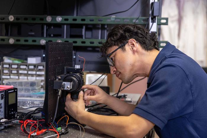
In a groundbreaking development at the University of Arizona Comprehensive Cancer Center, researcher Dongkyun Kang, PhD, is spearheading the creation of an innovative confocal microscope designed to detect chemotherapy-induced peripheral neuropathy (CIPN) earlier and more precisely than ever before. This noninvasive technology aims to revolutionize how clinicians diagnose and monitor this debilitating side effect, steering the medical community toward objective, quantitative biomarkers that could transform patient care. Backed by a substantial $2.4 million grant from the National Cancer Institute, Kang’s project promises to open new doors for personalized approaches to managing neuropathic symptoms linked to cancer therapy.
Chemotherapy-induced peripheral neuropathy is an often overlooked but profoundly impactful complication afflicting cancer patients undergoing treatment. Manifesting as numbness, weakness, and pain predominantly in the extremities, CIPN severely diminishes patients’ quality of life. The underlying pathology involves damage to peripheral nerves resulting from neurotoxic chemotherapeutic agents, presenting both diagnostic challenges and therapeutic dilemmas. Clinicians traditionally rely on subjective symptom reports and qualitative assessments, which are inherently limited and inconsistent, underscoring the urgent need for advanced diagnostic tools with high specificity and sensitivity.
The novel confocal microscope being developed under Kang’s leadership leverages sophisticated optical imaging techniques to noninvasively visualize nerve endings in the skin, focusing specifically on Meissner corpuscles. These specialized mechanoreceptors are critical for sensing light touch and low-frequency vibrations and are known to decrease in density in patients suffering from CIPN. By quantifying the number and condition of Meissner corpuscles through high-resolution confocal images, this technology aims to establish reliable biomarkers indicative of neuropathy progression or resolution.
Confocal microscopy, conventionally utilized in both research and clinical laboratories for cellular and subcellular imaging, offers unmatched optical sectioning capabilities and depth resolution. Kang’s approach enhances this modality by engineering a cost-effective, portable system that can be feasibly integrated into diverse clinical environments beyond traditional research settings. The innovation lies not only in the imaging hardware but also in the development of automated image analysis algorithms capable of identifying and quantifying nerve structures with precision, a significant stride towards the objective evaluation of peripheral neuropathies.
This initiative is also notable for its interdisciplinary collaboration, gathering expertise from the U of A James C. Wyant College of Optical Sciences, the College of Engineering’s Department of Biomedical Engineering, the U of A College of Medicine, and the Skin Cancer Institute. Furthermore, international partnerships with Guy’s and St. Thomas’ Hospital in London and the Memorial Sloan Kettering Cancer Center amplify the project’s clinical breadth and translational potential. Such a network of clinicians and scientists ensures that the microscope is rigorously tested across heterogeneous patient populations and cancer types, optimizing its diagnostic robustness.
One of the significant advantages of Kang’s low-cost confocal microscopy lies in its potential global accessibility. By circumventing the high expenses associated with conventional diagnostic imaging platforms, this technology could democratize CIPN monitoring, reaching community clinics and resource-limited settings worldwide. Early detection supported by quantitative biomarkers may enable oncologists to adjust chemotherapy regimens proactively, mitigate nerve damage, and tailor symptom management strategies, ultimately enhancing therapeutic outcomes and patient wellbeing.
The upcoming clinical trials endorsed by this grant will assess the confocal microscope’s efficacy in real-world oncology settings. Enrolling cancer patients actively undergoing chemotherapy, these studies aim to correlate confocal imaging findings with clinical symptomatology, electrophysiological measurements, and quality-of-life indices. Such comprehensive validation is crucial for regulatory approvals and establishing clinical guidelines for deploying this technology as a standard diagnostic tool for CIPN.
In addition to diagnostic applications, the confocal microscope may serve as a powerful research instrument for unraveling the pathophysiological mechanisms underpinning CIPN. By enabling longitudinal, high-resolution visualization of nerve fiber degeneration and regeneration, researchers can investigate therapeutic interventions’ efficacy and explore neuroprotective strategies. This could accelerate the discovery of new treatments while refining existing chemotherapy protocols to minimize neuropathic side effects.
Dongkyun Kang stresses the paradigm shift this tool represents in neuropathy diagnosis, moving away from subjective symptom checklists toward quantitative, image-based biomarkers. Such transformation aligns with the broader trend in precision medicine, harnessing advanced technologies to deliver individualized care grounded in measurable biological parameters. For patients, this could mean earlier interventions, reduced suffering, and improved functional outcomes.
Commenting on the significance of this work, Dan Theodorescu, MD, PhD, director of the University of Arizona Comprehensive Cancer Center, highlights the project’s potential for far-reaching impact on cancer care. He emphasizes how integrating optical engineering innovations into clinical oncology exemplifies the center’s commitment to precision prevention and therapy, reinforcing the importance of multidisciplinary research initiatives in tackling complex treatment-related complications.
Co-investigators playing pivotal roles in this endeavor include Clara Curiel-Lewandrowski, MD, a leading dermatologist and BIO5 Institute member, and Denise Roe, DrPH, an expert in biostatistics and bioinformatics. Their expertise ensures that clinical trial design, data analysis, and interpretation maintain the highest scientific standards. Collaborators from prominent institutions abroad lend additional clinical insight and facilitate cross-institutional knowledge exchange, further enriching the project’s scope.
Supported by the National Cancer Institute under award number 1R01CA301271-01, this research embodies the potential for significant advances in cancer survivorship care. As precision diagnostics continue to evolve, the confocal microscopy platform developed by Kang and colleagues stands as a promising beacon for transforming how chemotherapy-induced peripheral neuropathy is understood, detected, and ultimately managed at the bedside.
Subject of Research: Development of a noninvasive confocal microscope for early detection and quantification of chemotherapy-induced peripheral neuropathy biomarkers
Image Credits: Photo by Joshua Elz, University of Arizona Cancer Center
Keywords: Chemotherapy, Biomarkers, Medical treatments, Clinical medicine, Clinical neuroscience


